Fearon D T
Proc Natl Acad Sci U S A. 1979 Nov;76(11):5867-71. doi: 10.1073/pnas.76.11.5867.
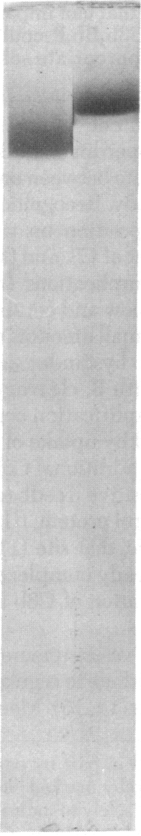
An activity that is inhibitory to the properdin-stabilized amplification C3 convertase (C3b,Bb,P) was solubilized from human erythrocyte (E(hu)) membranes by Nonidet P-40 and purified to homogeneity. The inhibitory membrane glycoprotein had an apparent M(r) of 1-1.2x10(6) on gel filtration in the presence of Nonidet P-40. On sodium dodecyl sulfate/polyacrylamide gel electrophoresis it presented a single stained band with an apparent M(r) of 205,000, with or without prior reduction of disulfides. The inhibitory protein of the E(hu) membrane produced a dose-related, first-order decay of C3b,Bb,P function on sheep erythrocytes (E(s)) and released (125)I-labeled Bb from these sites, indicating a mechanism of inhibition by decay-dissociation of the amplification C3 convertase. The 50% inhibitory dose of the E(hu) membrane protein was not altered by removal of sialic acid from the E(s) bearing C3b,Bb,P sites. E(hu) membrane protein also serves as a cofactor for C3b inactivator-induced cleavage of the alpha polypeptide chain of C3b. Thus, the inhibitory membrane protein can abrogate the activity of amplification convertase sites that have formed and also can prevent generation of such sites by augmenting irreversible inactivation of C3b.Discrimination between cells by the alternative complement pathway occurs after initial deposition of C3b and is related to the modulation by surface constituents of the capacity of bound C3b to function as a subunit of the amplification C3 convertase. The existence in the E(hu) membrane of a protein that can impair the functions of membrane-bound C3b and C3b,Bb,P could represent a molecular basis for preventing inappropriate self-recognition.
一种对备解素稳定的补体C3转化酶(C3b,Bb,P)具有抑制作用的活性物质,通过Nonidet P - 40从人红细胞(E(hu))膜中溶解出来,并纯化至同质。在存在Nonidet P - 40的情况下,通过凝胶过滤法测得该抑制性膜糖蛋白的表观分子量为1 - 1.2×10⁶。在十二烷基硫酸钠/聚丙烯酰胺凝胶电泳中,无论是否预先还原二硫键,它都呈现出一条表观分子量为205,000的单一染色带。E(hu)膜的抑制性蛋白对绵羊红细胞(E(s))上的C3b,Bb,P功能产生剂量相关的一级衰减,并从这些位点释放出¹²⁵I标记的Bb,这表明其抑制机制是通过扩增C3转化酶的衰变解离。从带有C3b,Bb,P位点的E(s)上去除唾液酸后,E(hu)膜蛋白的50%抑制剂量并未改变。E(hu)膜蛋白还可作为C3b灭活剂诱导C3bα多肽链裂解的辅因子。因此,这种抑制性膜蛋白既能消除已形成的扩增转化酶位点的活性,又能通过增强C3b的不可逆失活来阻止此类位点的产生。补体替代途径对细胞的识别是在C3b初始沉积之后发生的,并且与表面成分对结合的C3b作为扩增C3转化酶亚基功能的调节能力有关。E(hu)膜中存在一种能够损害膜结合C3b和C3b,Bb,P功能的蛋白质,这可能是防止不适当自我识别的分子基础。