Le Binh Duong, Kang Donggu, Yun Seokhwan, Jeong Young Hun, Kwak Jong-Young, Yoon Sik, Jin Songwan
Department of Advanced Convergence Technology, Korea Polytechnic Univsersity, Siheung-si 15073, Gyoenggi-do, Korea.
Department of Mechanical System Engineering, Korea Polytechnic Univsersity, Siheung-si 15073, Gyoenggi-do, Korea.
Nanomaterials (Basel). 2018 Jan 25;8(2):64. doi: 10.3390/nano8020064.
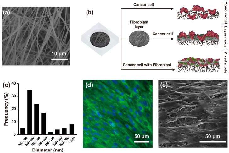
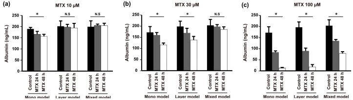
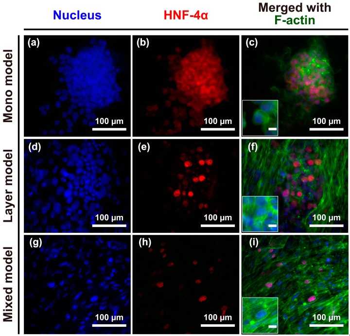
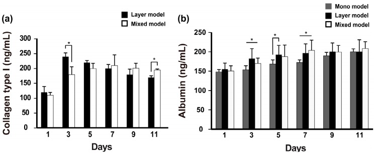
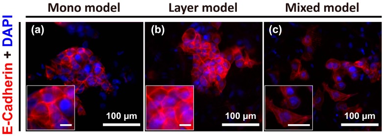
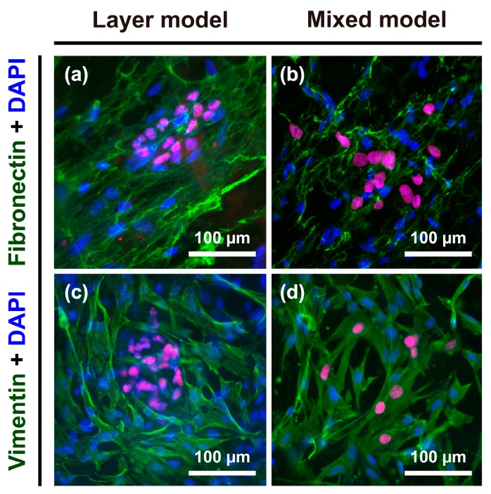
Three-dimensional (3D) in vitro tissue or organ models can effectively mimic the complex microenvironment of many types of human tissues for medical applications. Unfortunately, development of 3D cancer models, which involve cancer/stromal cells in a 3D environment, has remained elusive due to the extreme complexity of the tumor microenvironment (TME) and the stepwise progression of human cancer. Here, we developed hepatocellular carcinoma (HCC) models, which consist of fibroblasts as stromal cells, HCC cells, and a nanofibrous membrane to mimic the complex TME. The 3D HCC models were fabricated using three distinct culture methods: cancer cells grown directly on the nanofibrous membrane (mono model), fibroblasts covering the nanofibrous membrane (layer model), and both cancer cells and fibroblasts grown on the nanofibrous membrane (mixed model). Interestingly, the mono model and layer model showed similar tissue structures, whereas the mixed model resulted in phenotypic changes to the cancer cells. Further analysis demonstrated that the mixed models promoted the expression of fibronectin and vimentin, and showed higher resistance to anticancer drugs compared with the other models. Thus, our 3D HCC model could be utilized for testing efficient anticancer therapies at various stages of cancer, with potential application to different tumor types.
三维(3D)体外组织或器官模型能够有效地模拟多种人体组织的复杂微环境,以用于医学应用。不幸的是,由于肿瘤微环境(TME)的极端复杂性和人类癌症的逐步进展,涉及在三维环境中的癌症/基质细胞的三维癌症模型的开发仍然难以实现。在这里,我们开发了肝细胞癌(HCC)模型,该模型由作为基质细胞的成纤维细胞、肝癌细胞和纳米纤维膜组成,以模拟复杂的肿瘤微环境。使用三种不同的培养方法构建了三维肝癌模型:癌细胞直接生长在纳米纤维膜上(单模型)、成纤维细胞覆盖纳米纤维膜(层模型)以及癌细胞和成纤维细胞都生长在纳米纤维膜上(混合模型)。有趣的是,单模型和层模型显示出相似的组织结构,而混合模型导致癌细胞发生表型变化。进一步分析表明,混合模型促进了纤连蛋白和波形蛋白的表达,并且与其他模型相比,对抗癌药物表现出更高的抗性。因此,我们的三维肝癌模型可用于在癌症的各个阶段测试有效的抗癌疗法,并有可能应用于不同的肿瘤类型。