• 文献检索
  • 文档翻译
  • 深度研究
  • 学术资讯
  • Suppr Zotero 插件Zotero 插件
  • 邀请有礼
  • 套餐&价格
  • 历史记录
应用&插件
Suppr Zotero 插件Zotero 插件浏览器插件Mac 客户端Windows 客户端微信小程序
定价
高级版会员购买积分包购买API积分包
服务
文献检索文档翻译深度研究API 文档MCP 服务
关于我们
关于 Suppr公司介绍联系我们用户协议隐私条款
关注我们

Suppr 超能文献

核心技术专利:CN118964589B侵权必究
粤ICP备2023148730 号-1Suppr @ 2026

文献检索

告别复杂PubMed语法,用中文像聊天一样搜索,搜遍4000万医学文献。AI智能推荐,让科研检索更轻松。

立即免费搜索

文件翻译

保留排版,准确专业,支持PDF/Word/PPT等文件格式,支持 12+语言互译。

免费翻译文档

深度研究

AI帮你快速写综述,25分钟生成高质量综述,智能提取关键信息,辅助科研写作。

立即免费体验

脊椎动物视网膜中的神经元迁移与分层

Neuronal Migration and Lamination in the Vertebrate Retina.

作者信息

Amini Rana, Rocha-Martins Mauricio, Norden Caren

机构信息

Max Planck Institute of Molecular Cell Biology and Genetics, Dresden, Germany.

出版信息

Front Neurosci. 2018 Jan 9;11:742. doi: 10.3389/fnins.2017.00742. eCollection 2017.

DOI:10.3389/fnins.2017.00742
PMID:29375289
原文链接:https://pmc.ncbi.nlm.nih.gov/articles/PMC5767219/
Abstract

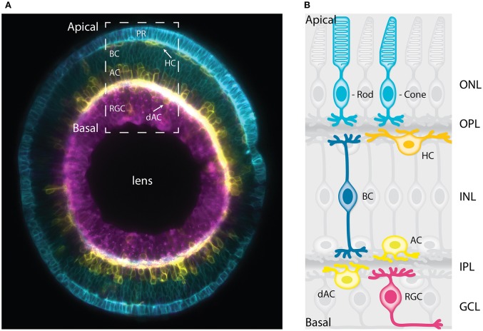
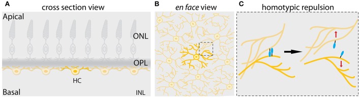
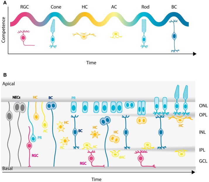
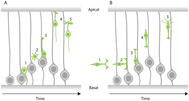
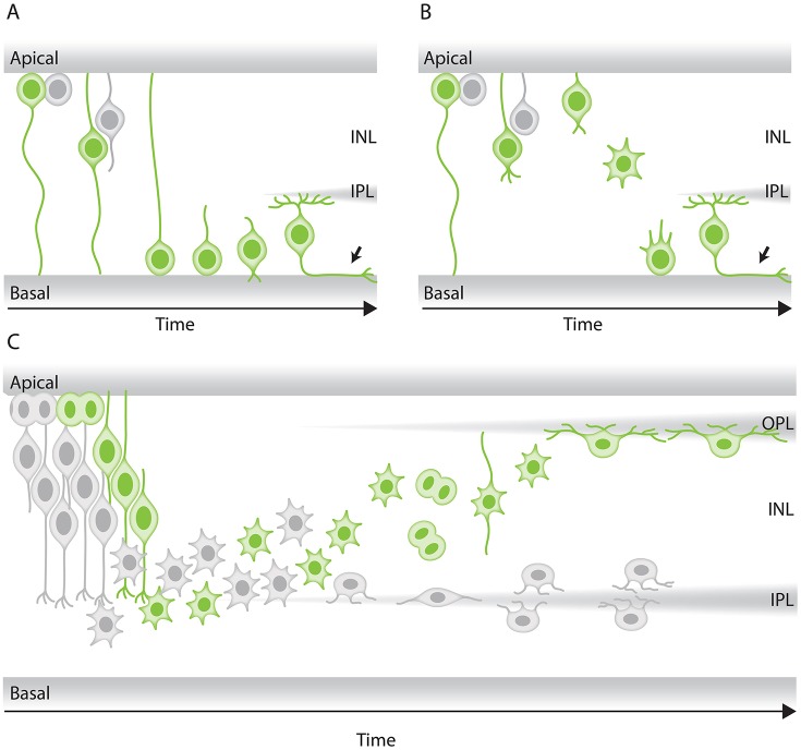
In the retina, like in most other brain regions, developing neurons are arranged into distinct layers giving the mature tissue its stratified appearance. This process needs to be highly controlled and orchestrated, as neuronal layering defects lead to impaired retinal function. To achieve successful neuronal layering and lamination in the retina and beyond, three main developmental steps need to be executed: First, the correct type of neuron has to be generated at a precise developmental time. Second, as most retinal neurons are born away from the position at which they later function, newborn neurons have to move to their final layer within the developing tissue, a process also termed neuronal lamination. Third, these neurons need to connect to their correct synaptic partners. Here, we discuss neuronal migration and lamination in the vertebrate retina and summarize our knowledge on these aspects of retinal development. We give an overview of how lamination emerges and discuss the different modes of neuronal translocation that occur during retinogenesis and what we know about the cell biological machineries driving them. In addition, retinal mosaics and their importance for correct retinal function are examined. We close by stating the open questions and future directions in this exciting field.

摘要

在视网膜中,如同在大多数其他脑区一样,发育中的神经元排列成不同的层,使成熟组织呈现出分层外观。这个过程需要高度控制和精心编排,因为神经元分层缺陷会导致视网膜功能受损。为了在视网膜及其他部位成功实现神经元分层和板层形成,需要执行三个主要的发育步骤:第一,必须在精确的发育时间产生正确类型的神经元。第二,由于大多数视网膜神经元在远离其后来发挥功能的位置产生,新生神经元必须迁移到发育组织内的最终层,这个过程也称为神经元板层形成。第三,这些神经元需要与正确的突触伙伴建立连接。在此,我们讨论脊椎动物视网膜中的神经元迁移和板层形成,并总结我们在视网膜发育这些方面的知识。我们概述板层形成是如何出现的,讨论视网膜发生过程中发生的神经元移位的不同模式,以及我们对驱动它们的细胞生物学机制的了解。此外,还研究了视网膜镶嵌及其对正确视网膜功能的重要性。最后,我们阐述了这个令人兴奋的领域中尚未解决的问题和未来的研究方向。

https://cdn.ncbi.nlm.nih.gov/pmc/blobs/4c3e/5767219/4e1fd8c314fd/fnins-11-00742-g0005.jpg
https://cdn.ncbi.nlm.nih.gov/pmc/blobs/4c3e/5767219/b3e870003d20/fnins-11-00742-g0001.jpg
https://cdn.ncbi.nlm.nih.gov/pmc/blobs/4c3e/5767219/e2c4307939ce/fnins-11-00742-g0002.jpg
https://cdn.ncbi.nlm.nih.gov/pmc/blobs/4c3e/5767219/7863366c402a/fnins-11-00742-g0003.jpg
https://cdn.ncbi.nlm.nih.gov/pmc/blobs/4c3e/5767219/484fbd80c95e/fnins-11-00742-g0004.jpg
https://cdn.ncbi.nlm.nih.gov/pmc/blobs/4c3e/5767219/4e1fd8c314fd/fnins-11-00742-g0005.jpg

相似文献

1
Neuronal Migration and Lamination in the Vertebrate Retina.脊椎动物视网膜中的神经元迁移与分层
Front Neurosci. 2018 Jan 9;11:742. doi: 10.3389/fnins.2017.00742. eCollection 2017.
2
Stochastic single cell migration leads to robust horizontal cell layer formation in the vertebrate retina.随机单细胞迁移导致脊椎动物视网膜中水平细胞层的稳健形成。
Development. 2019 May 24;146(12):dev173450. doi: 10.1242/dev.173450.
3
Independent modes of ganglion cell translocation ensure correct lamination of the zebrafish retina.神经节细胞移位的独立模式确保斑马鱼视网膜的正确分层。
J Cell Biol. 2016 Oct 24;215(2):259-275. doi: 10.1083/jcb.201604095.
4
Retinal neuroblast migration and ganglion cell layer organization require the cytoskeletal-interacting protein Mllt11.视网膜神经母细胞迁移和节细胞层组织需要细胞骨架相互作用蛋白 Mllt11。
Dev Dyn. 2023 Feb;252(2):305-319. doi: 10.1002/dvdy.540. Epub 2022 Oct 4.
5
Inhibitory neuron migration and IPL formation in the developing zebrafish retina.发育中的斑马鱼视网膜中抑制性神经元的迁移和内网状层的形成。
Development. 2015 Aug 1;142(15):2665-77. doi: 10.1242/dev.122473. Epub 2015 Jun 26.
6
is required for the integrity of the photoreceptor layer in the zebrafish retina.对于斑马鱼视网膜中光感受器层的完整性是必需的。
Biol Open. 2019 Apr 23;8(4):bio041830. doi: 10.1242/bio.041830.
7
Development of the lateral geniculate nucleus: interactions between retinal afferent, cytoarchitectonic, and glial cell process lamination in ferrets and tree shrews.外侧膝状体核的发育:雪貂和树鼩中视网膜传入、细胞构筑和胶质细胞突起分层之间的相互作用。
J Comp Neurol. 1990 Aug 1;298(1):113-28. doi: 10.1002/cne.902980109.
8
βII-Spectrin Is Required for Synaptic Positioning during Retinal Development.βII- spectrin 在视网膜发育过程中对于突触定位是必需的。
J Neurosci. 2023 Jul 19;43(29):5277-5289. doi: 10.1523/JNEUROSCI.0063-23.2023. Epub 2023 Jun 27.
9
A Fish Eye View: Retinal Morphogenesis from Optic Cup to Neuronal Lamination.鱼眼视角:从视杯到神经元分层的视网膜形态发生
Annu Rev Cell Dev Biol. 2023 Oct 16;39:175-196. doi: 10.1146/annurev-cellbio-012023-013036. Epub 2023 Jul 7.
10
Patterning the vertebrate retina: the early appearance of retinal mosaics.构建脊椎动物视网膜:视网膜镶嵌的早期出现。
Semin Cell Dev Biol. 1998 Jun;9(3):279-84. doi: 10.1006/scdb.1998.0233.

引用本文的文献

1
Timely neurogenesis drives the transition from nematic to crystalline nuclear packing during retinal morphogenesis.在视网膜形态发生过程中,及时的神经发生驱动了从向列型到晶体状核排列的转变。
Sci Adv. 2025 May 9;11(19):eadu6843. doi: 10.1126/sciadv.adu6843.
2
Influences of lead-based perovskite nanoparticles exposure on early development of human retina.基于铅的钙钛矿纳米颗粒暴露对人类视网膜早期发育的影响。
J Nanobiotechnology. 2025 Feb 26;23(1):144. doi: 10.1186/s12951-025-03245-w.
3
Ultrastructural imaging biomarkers in diabetic macular edema: A major review.

本文引用的文献

1
Phototoxicity in live fluorescence microscopy, and how to avoid it.活细胞荧光显微镜检查中的光毒性及其规避方法。
Bioessays. 2017 Aug;39(8). doi: 10.1002/bies.201700003.
2
The Functioning of a Cortex without Layers.无层皮质的功能
Front Neuroanat. 2017 Jul 12;11:54. doi: 10.3389/fnana.2017.00054. eCollection 2017.
3
Disaggregation and Reaggregation of Zebrafish Retinal Cells for the Analysis of Neuronal Layering.用于神经元分层分析的斑马鱼视网膜细胞的解离与重聚集
糖尿病性黄斑水肿中的超微结构成像生物标志物:一项主要综述。
Indian J Ophthalmol. 2025 Jan 1;73(Suppl 1):S7-S23. doi: 10.4103/IJO.IJO_878_24. Epub 2024 Dec 24.
4
Connexin Expression Is Altered in the Eye Development of Mice: A Preliminary Study.缝隙连接蛋白在小鼠眼部发育过程中的表达变化:初步研究。
Biomolecules. 2024 Sep 19;14(9):1174. doi: 10.3390/biom14091174.
5
The intracellular domain of Sema6A is essential for development of the zebrafish retina.Sema6A的细胞内结构域对于斑马鱼视网膜的发育至关重要。
J Cell Sci. 2024 Jul 15;137(14). doi: 10.1242/jcs.261469. Epub 2024 Jul 25.
6
Decreased CREB phosphorylation impairs embryonic retinal neurogenesis in the mouse model of Ocular albinism.在眼白化病小鼠模型中,CREB磷酸化水平降低会损害胚胎视网膜神经发生。
bioRxiv. 2024 May 14:2024.05.14.594013. doi: 10.1101/2024.05.14.594013.
7
Advances in Extraction Protocols, Degradation Methods, and Bioactivities of Proanthocyanidins.原花青素提取方法、降解方法和生物活性的研究进展。
Molecules. 2024 May 7;29(10):2179. doi: 10.3390/molecules29102179.
8
Retina organoids: Window into the biophysics of neuronal systems.视网膜类器官:洞察神经元系统生物物理学的窗口。
Biophys Rev (Melville). 2022 Jan 18;3(1):011302. doi: 10.1063/5.0077014. eCollection 2022 Mar.
9
Transplanted human photoreceptors transfer cytoplasmic material but not to the recipient mouse retina.移植的人眼感光细胞会转移细胞质物质,但不会转移到受者小鼠的视网膜中。
Stem Cell Res Ther. 2024 Mar 14;15(1):79. doi: 10.1186/s13287-024-03679-3.
10
Deficiency of copper responsive gene stmn4 induces retinal developmental defects.铜反应基因 stmn4 的缺乏会导致视网膜发育缺陷。
Cell Biol Toxicol. 2024 Jan 22;40(1):2. doi: 10.1007/s10565-024-09847-8.
Methods Mol Biol. 2019;1576:255-271. doi: 10.1007/7651_2017_46.
4
Concerted action of neuroepithelial basal shrinkage and active epithelial migration ensures efficient optic cup morphogenesis.神经上皮基底收缩和活跃的上皮迁移的协同作用确保了有效的视杯形态发生。
Elife. 2017 Apr 4;6:e22689. doi: 10.7554/eLife.22689.
5
Uncoupling of neurogenesis and differentiation during retinal development.视网膜发育过程中神经发生与分化的解偶联。
EMBO J. 2017 May 2;36(9):1134-1146. doi: 10.15252/embj.201694230. Epub 2017 Mar 3.
6
Self-organising aggregates of zebrafish retinal cells for investigating mechanisms of neural lamination.用于研究神经分层机制的斑马鱼视网膜细胞自组织聚集体。
Development. 2017 Mar 15;144(6):1097-1106. doi: 10.1242/dev.142760. Epub 2017 Feb 7.
7
Independent modes of ganglion cell translocation ensure correct lamination of the zebrafish retina.神经节细胞移位的独立模式确保斑马鱼视网膜的正确分层。
J Cell Biol. 2016 Oct 24;215(2):259-275. doi: 10.1083/jcb.201604095.
8
Fat3 and Ena/VASP proteins influence the emergence of asymmetric cell morphology in the developing retina.Fat3和Ena/VASP蛋白影响发育中视网膜不对称细胞形态的出现。
Development. 2016 Jun 15;143(12):2172-82. doi: 10.1242/dev.133678. Epub 2016 Apr 27.
9
Characterization of primary cilia during the differentiation of retinal ganglion cells in the zebrafish.斑马鱼视网膜神经节细胞分化过程中初级纤毛的特征分析。
Neural Dev. 2016 Apr 6;11:10. doi: 10.1186/s13064-016-0064-z.
10
Identity of neocortical layer 4 neurons is specified through correct positioning into the cortex.新皮层第4层神经元的身份是通过正确定位到皮层中而确定的。
Elife. 2016 Feb 12;5:e10907. doi: 10.7554/eLife.10907.