Lee Yeon-Hee, Lee Kyung Mi, Auh Q-Schick, Hong Jyung-Pyo
Orofacial Pain and Oral Medicine, Kyung Hee University Dental Hospital, Seoul, South Korea.
Radiology, Kyung Hee University College of Medicine, Kyung Hee University Hospital, Seoul, South Korea.
Front Neurol. 2018 Jan 9;8:725. doi: 10.3389/fneur.2017.00725. eCollection 2017.
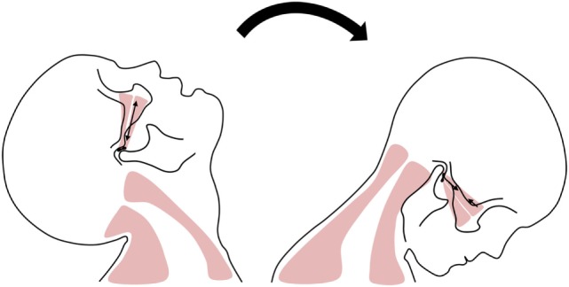
Whiplash injury can cause internal derangement of the temporomandibular joint (TMJ) and lead to temporomandibular disorders (TMDs). Our aim was to evaluate whether the initial clinical findings in TMD patients with whiplash injury are correlated with their magnetic resonance imaging (MRI) characteristics.
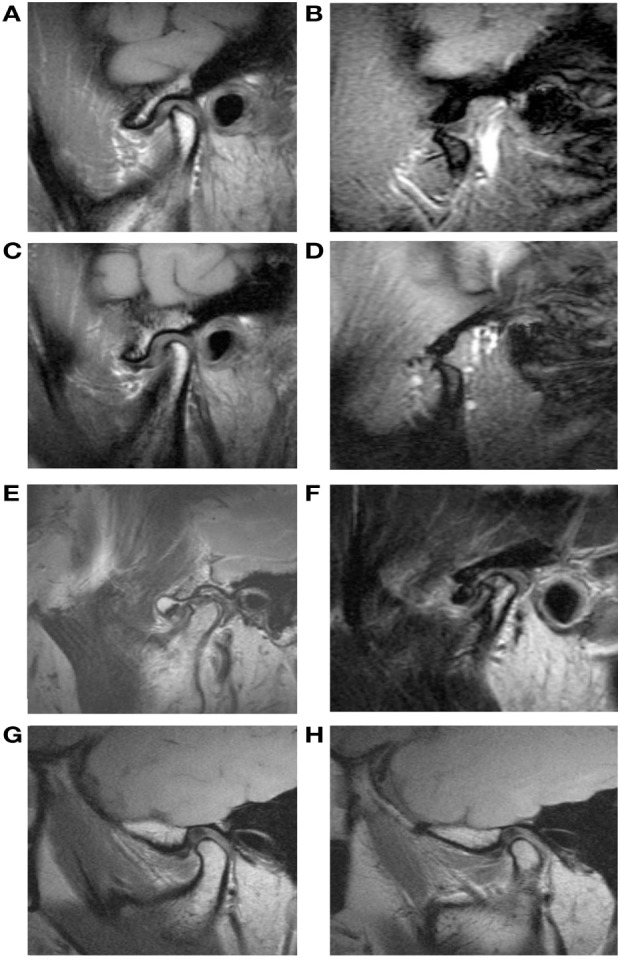
This case-control study involved 219 patients (135 women, 84 men; mean age: 37.84 years) who visited our orofacial pain clinic with TMD; TMD was diagnosed using the diagnostic criteria for TMD Axis I. Patients were categorized into three groups based on the presence and type of macrotrauma: in the "wTMD" group, patients had suffered whiplash injury; patients in the "pTMD" group had post-traumatic TMD; the "iTMD" group comprised patients who had presented with TMD symptoms and had sustained no macrotrauma. We investigated the presence of disk displacement, effusion, disk deformity, and condylar degeneration, and changes in the lateral pterygoid muscle (LPM). To evaluate the severity of TMD pain and objectively analyze symptoms, we used a visual analog scale (VAS), palpation index (PI), neck PI, dysfunction index, and craniomandibular index (CMI).
The VAS scores, and the severity indexes of the TMD including PI, neck PI, and CMI were highest in the wTMD patients. Atrophy of the LPM was most commonly seen in the wTMD group, as was disk deformity. In wTMD patients only, VAS score was significantly correlated with stress; it was correlated with headache in wTMD and iTMD patients. The clinical symptoms of TMD were not correlated with MRI findings in the wTMD group. However, alterations in the LPM were strongly correlated with disk displacement.
If clinicians recognize alterations in the LPM and disk displacement in the TMJ, they will better understand the clinical symptoms and pathophysiology of TMD with whiplash injury. Whiplash injury may lead to TMD different mechanisms from other macrotraumas.
挥鞭样损伤可导致颞下颌关节(TMJ)内部紊乱并引发颞下颌关节紊乱病(TMD)。我们的目的是评估伴有挥鞭样损伤的TMD患者的初始临床发现是否与其磁共振成像(MRI)特征相关。
本病例对照研究纳入了219例因TMD前来我们口腔颌面疼痛诊所就诊的患者(135例女性,84例男性;平均年龄:37.84岁);TMD采用TMD轴I诊断标准进行诊断。根据是否存在宏观创伤及其类型,患者被分为三组:“wTMD”组患者遭受过挥鞭样损伤;“pTMD”组患者患有创伤后TMD;“iTMD”组包括出现TMD症状但未遭受宏观创伤的患者。我们调查了盘移位、积液、盘变形和髁突退变的情况,以及翼外肌(LPM)的变化。为评估TMD疼痛的严重程度并客观分析症状,我们使用了视觉模拟量表(VAS)、触诊指数(PI)、颈部PI、功能障碍指数和颅下颌指数(CMI)。
wTMD患者的VAS评分以及包括PI、颈部PI和CMI在内的TMD严重程度指数最高。LPM萎缩在wTMD组中最为常见,盘变形也是如此。仅在wTMD患者中,VAS评分与压力显著相关;在wTMD和iTMD患者中,它与头痛相关。wTMD组中TMD的临床症状与MRI表现不相关。然而,LPM的改变与盘移位密切相关。
如果临床医生认识到TMJ中LPM的改变和盘移位,他们将更好地理解伴有挥鞭样损伤的TMD的临床症状和病理生理学。挥鞭样损伤可能通过与其他宏观创伤不同的机制导致TMD。