Tucholka Alan, Grau-Rivera Oriol, Falcon Carles, Rami Lorena, Sánchez-Valle Raquel, Lladó Albert, Gispert Juan Domingo, Molinuevo José Luis
Barcelonaβeta Brain Research Center, Pasqual Maragall Foundation, Barcelona, Spain.
Alzheimer's Disease and Other Cognitive Disorders Unit, Hospital Clínic, Institut d'Investigacions Biomèdiques August Pi i Sunyer (IDIBAPS), Barcelona, Spain.
J Alzheimers Dis. 2018;61(4):1575-1587. doi: 10.3233/JAD-170553.
Gray matter changes associated with the progression of Alzheimer's disease (AD) have been thoroughly studied. However, alterations in white matter tracts have received less attention, particularly during early or preclinical stages of the disease.
To identify the structural connectivity changes across the AD continuum.
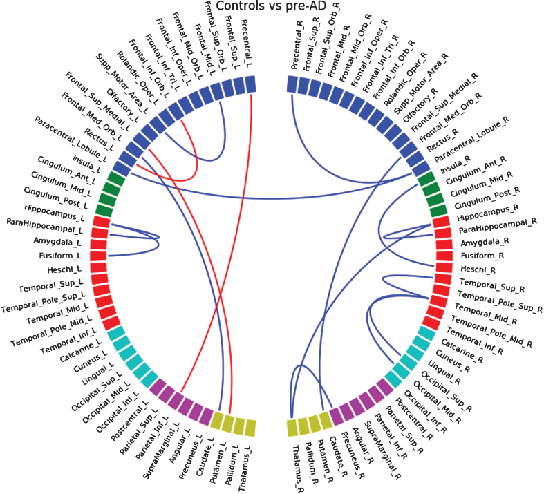
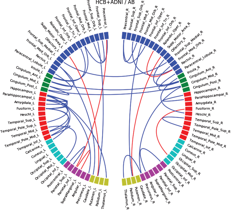
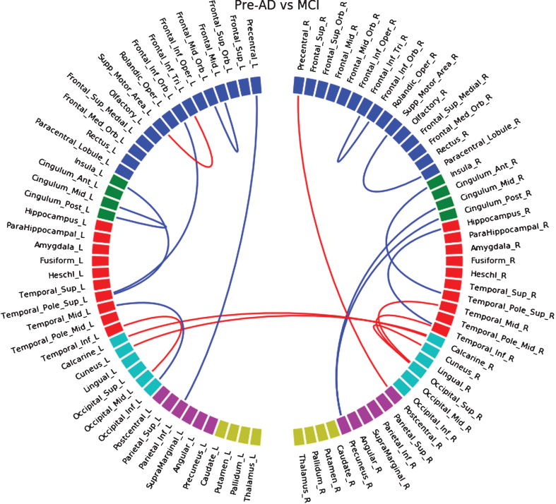
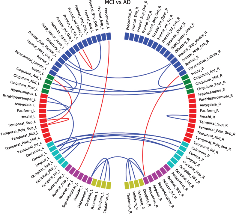
We performed probabilistic tractography in a total of 183 subjects on two independent samples that include control (n = 68) and preclinical AD individuals (n = 28), patients diagnosed with mild cognitive impairment (MCI) due to AD (n = 44), and AD patients (n = 43). We compared the connectivity between groups, and with CSF Aβ42 and tau biomarkers.
We observed disconnections in preclinical individuals, mainly located in the temporal lobe. This pattern of disconnection spread to the parietal and frontal lobes at the MCI stage and involved almost all the brain in AD. These findings were not driven by gray matter atrophy.
Using tractography, we were able to identify white matter changes between subsequent disease stages and, notably, also in preclinical AD. Therefore, this method may be useful for detecting early and specific brain structural changes during preclinical AD stage.
与阿尔茨海默病(AD)进展相关的灰质变化已得到充分研究。然而,白质束的改变受到的关注较少,尤其是在疾病的早期或临床前期阶段。
确定AD连续体中的结构连接性变化。
我们在总共183名受试者的两个独立样本上进行了概率纤维束成像,样本包括对照组(n = 68)和临床前期AD个体(n = 28)、因AD诊断为轻度认知障碍(MCI)的患者(n = 44)以及AD患者(n = 43)。我们比较了各组之间的连接性,以及与脑脊液Aβ42和tau生物标志物的连接性。
我们在临床前期个体中观察到了连接中断,主要位于颞叶。这种连接中断模式在MCI阶段扩散到顶叶和额叶,并在AD中累及几乎整个大脑。这些发现并非由灰质萎缩所致。
使用纤维束成像,我们能够识别后续疾病阶段之间的白质变化,特别是在临床前期AD中。因此,这种方法可能有助于在临床前期AD阶段检测早期和特定的脑结构变化。