Chanani Ankit, Adhikari Haridas Das
Department of Conservative Dentistry and Endodontics, Dr. R. Ahmed Dental College and Hospital, Kolkata, West Bengal, India.
J Conserv Dent. 2017 Sep-Oct;20(5):326-331. doi: 10.4103/JCD.JCD_124_17.
Differential diagnosis of periapical cysts and granulomas is required as their treatment modalities are different.
The aim of this study was to evaluate the efficacy of cone beam computed tomography (CBCT) in the differential diagnosis of periapical cysts from granulomas.
A single-centered observational study was carried out in the Department of Conservative Dentistry and Endodontics, Dr. R. Ahmed Dental College and Hospital, using CBCT and dental operating microscope.
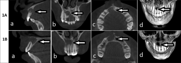
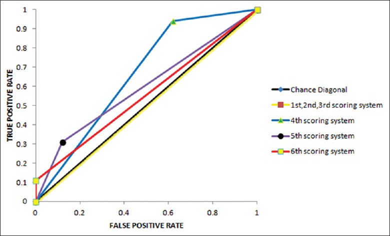
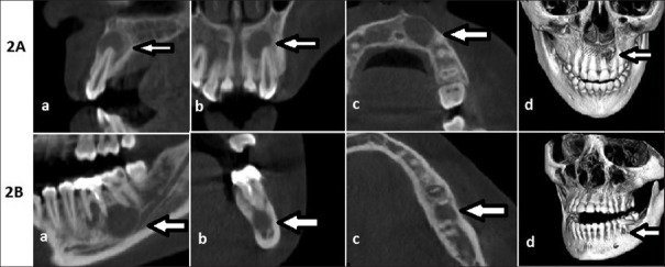
Forty-five lesions were analyzed using CBCT scans. One evaluator analyzed each CBCT scan for the presence of the following six characteristic radiological features: cyst like-location, shape, periphery, internal structure, effect on the surrounding structures, and cortical plate perforation. Another independent evaluator analyzed the CBCT scans. This process was repeated after 6 months, and inter- and intrarater reliability of CBCT diagnoses was evaluated. Periapical surgeries were performed and tissue samples were obtained for histopathological analysis. To evaluate the efficacy, CBCT diagnoses were compared with histopathological diagnoses, and six receiver operating characteristic (ROC) curve analyses were conducted.
ROC curve, Cronbach's alpha (α) test, and Cohen Kappa (κ) test were used for statistical analysis.
Both inter- and intrarater reliability were excellent (α = 0.94, κ = 0.75 and 0.77, respectively). ROC curve with regard to ≥4 positive findings revealed the highest area under curve (0.66).
CBCT is moderately accurate in the differential diagnosis of periapical cysts and granulomas.
由于根尖囊肿和肉芽肿的治疗方式不同,因此需要对它们进行鉴别诊断。
本研究的目的是评估锥形束计算机断层扫描(CBCT)在根尖囊肿与肉芽肿鉴别诊断中的效能。
在R. Ahmed博士牙科学院及医院的保守牙科与牙髓病科开展了一项单中心观察性研究,使用CBCT和牙科手术显微镜。
使用CBCT扫描分析45个病变。一名评估者分析每个CBCT扫描,以确定是否存在以下六种特征性放射学表现:囊肿样位置、形状、边缘、内部结构、对周围结构的影响以及皮质板穿孔。另一名独立评估者分析CBCT扫描。6个月后重复此过程,并评估CBCT诊断的评估者间和评估者内可靠性。进行根尖手术并获取组织样本进行组织病理学分析。为评估效能,将CBCT诊断与组织病理学诊断进行比较,并进行六项受试者工作特征(ROC)曲线分析。
使用ROC曲线、Cronbach's alpha(α)检验和Cohen Kappa(κ)检验进行统计分析。
评估者间和评估者内可靠性均极佳(α分别为0.94,κ分别为0.75和0.77)。关于≥4项阳性发现的ROC曲线显示曲线下面积最高(0.66)。
CBCT在根尖囊肿和肉芽肿的鉴别诊断中具有中等准确性。