Discipline of Restorative Dentistry and Endodontics, Research Center TADERP, Faculty of Dental Medicine, "Victor Babes" University of Medicine and Pharmacy, 300041 Timisoara, Romania.
Discipline of Pedodontics, Pediatric Dentistry Research Center, Faculty of Dental Medicine, "Victor Babes" University of Medicine and Pharmacy, 300041 Timisoara, Romania.
Medicina (Kaunas). 2021 May 14;57(5):497. doi: 10.3390/medicina57050497.
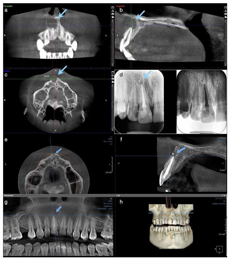
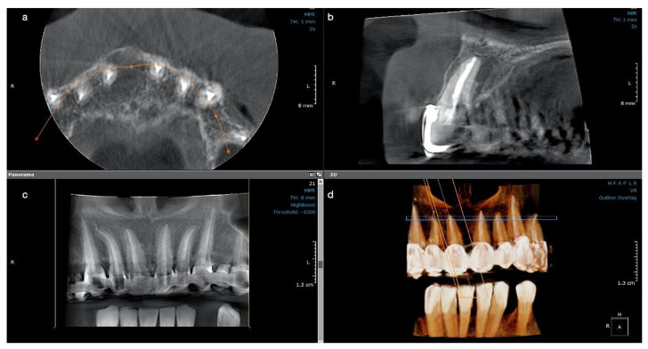
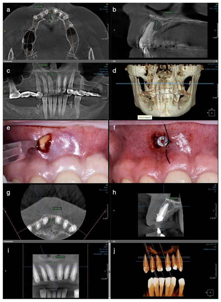
: Periapical cystic lesions are a pathology frequently addressed to endodontic specialists. Although their therapy is still not standardized, the treatment should be as conservative as possible and by endodontic means, as they are lesions of endodontic origin. The present case report describes two cases of upper central incisors with large cyst-like periapical lesions, and their one-year follow up. : Endodontic orthograde treatment was performed under copious irrigation with sodium hypochlorite, in association with calcium hydroxide as an intra-canal medication for both teeth. Root canal filling was achieved in a separate appointment using the continuous wave of condensation technique. A decompression procedure was used in association with endodontic therapy in the second case to reduce the pressure inside the cystic lesion and to allow its drainage, and only because the root canal could not be dried three weeks after medication. Initial cone beam computed tomography (CBCT) investigations, as well as at the one-year follow up, were used to compare the evolution of the lesion. : Both cases had a favorable outcome. New bone formation in the periapical region and complete resolution of the lesion was observed at the one-year control in the first case. In the second case, although the lesion was still not completely healed at 12 months, a significant reduction in its size could be observed, showing active signs of healing. : Endodontic treatment is the first choice option in the management of teeth with pulpal necrosis and large periapical cystic-like lesions. Decompression is the only surgical procedure recommended when the canals cannot be dried and obturated. Large surgical interventions are unnecessary in cases where endodontic treatment can be performed.
: 根尖周囊肿是经常需要牙髓专科医生处理的一种病症。尽管其治疗方法尚未标准化,但治疗应尽可能保守,并通过牙髓治疗手段进行,因为这些病变是牙髓来源的。本病例报告描述了 2 例上颌中切牙伴有大的囊肿样根尖周病变及其 1 年的随访情况。: 采用大量次氯酸钠冲洗,联合氢氧化钙作为根管内药物,对上颌中切牙进行根管治疗。在另一时间段,使用连续波冷凝技术进行根管充填。在第二例中,由于根管治疗后 3 周仍无法干燥,联合牙髓治疗使用减压术以降低囊腔内部压力并允许其引流。初始锥形束 CT(CBCT)检查以及 1 年随访用于比较病变的演变。: 这两例均有良好的预后。在第 1 例中,1 年后可见根尖周区域有新骨形成,病变完全消退。在第 2 例中,尽管在 12 个月时病变仍未完全愈合,但可以观察到其明显缩小,显示出活跃的愈合迹象。: 对于牙髓坏死和大的囊肿样根尖周病变的患牙,牙髓治疗是首选治疗方法。当根管无法干燥和封闭时,减压术是唯一推荐的手术方法。对于可以进行牙髓治疗的病例,无需进行大型手术干预。