Mezzanotte Laura, Blankevoort Vicky, Löwik Clemens W G M, Kaijzel Eric L
Department of Radiology, Experimental Molecular Imaging, Leiden University Medical Center, Albinusdreef 2, 2333 ZA, Leiden, The Netherlands.
Anal Bioanal Chem. 2014 Sep;406(23):5727-34. doi: 10.1007/s00216-014-7917-2. Epub 2014 Jun 24.
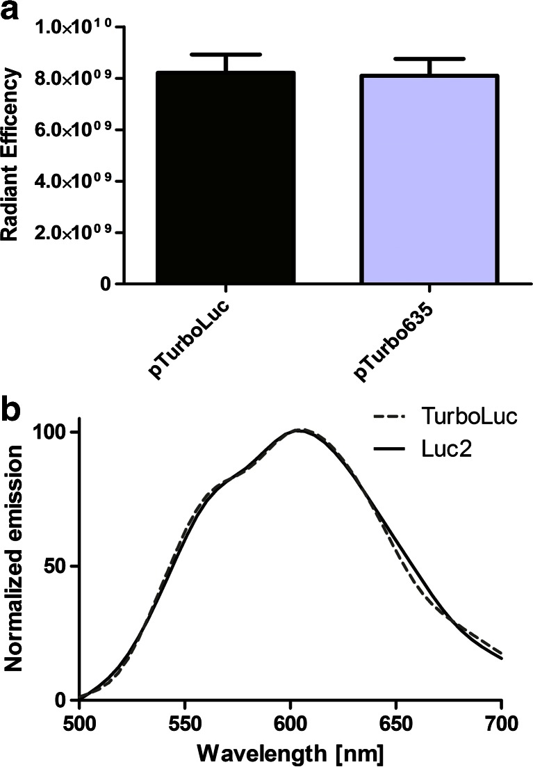
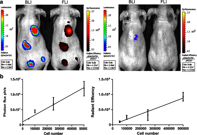
Fluorescence and bioluminescence imaging have different advantages and disadvantages depending on the application. Bioluminescence imaging is now the most sensitive optical technique for tracking cells, promoter activity studies, or for longitudinal in vivo preclinical studies. Far-red and near-infrared fluorescence imaging have the advantage of being suitable for both ex vivo and in vivo analysis and have translational potential, thanks to the availability of very sensitive imaging instrumentation. Here, we report the development and validation of a new luciferase fusion reporter generated by the fusion of the firefly luciferase Luc2 to the far-red fluorescent protein TurboFP635 by a 14-amino acid linker peptide. Expression of the fusion protein, named TurboLuc, was analyzed in human embryonic kidney cells, (HEK)-293 cells, via Western blot analysis, fluorescence microscopy, and in vivo optical imaging. The created fusion protein maintained the characteristics of the original bioluminescent and fluorescent protein and showed no toxicity when expressed in living cells. To assess the sensitivity of the reporter for in vivo imaging, transfected cells were subcutaneously injected in animals. Detection limits of cells were 5 × 10(3) and 5 × 10(4) cells for bioluminescent and fluorescent imaging, respectively. In addition, hydrodynamics-based in vivo gene delivery using a minicircle vector expressing TurboLuc allowed for the analysis of luminescent signals over time in deep tissue. Bioluminescence could be monitored for over 30 days in the liver of animals. In conclusion, TurboLuc combines the advantages of both bioluminescence and fluorescence and allows for highly sensitive optical imaging ranging from single-cell analysis to in vivo whole-body bioluminescence imaging.
根据应用的不同,荧光成像和生物发光成像各有优缺点。生物发光成像目前是用于追踪细胞、启动子活性研究或纵向体内临床前研究的最灵敏的光学技术。远红光和近红外荧光成像具有适用于离体和体内分析的优点,并且由于有非常灵敏的成像仪器,因此具有转化潜力。在此,我们报告了一种新的荧光素酶融合报告基因的开发和验证,该报告基因是通过萤火虫荧光素酶Luc2与远红光荧光蛋白TurboFP635通过一个14个氨基酸的连接肽融合而成。通过蛋白质免疫印迹分析、荧光显微镜检查和体内光学成像,在人胚肾细胞(HEK)-293细胞中分析了名为TurboLuc的融合蛋白的表达。所产生的融合蛋白保留了原始生物发光蛋白和荧光蛋白的特性,并且在活细胞中表达时无毒性。为了评估该报告基因用于体内成像的灵敏度,将转染的细胞皮下注射到动物体内。生物发光成像和荧光成像检测细胞的下限分别为5×10³和5×10⁴个细胞。此外,使用表达TurboLuc的微小环载体基于流体动力学的体内基因递送允许分析深部组织中随时间变化的发光信号。在动物肝脏中可以监测生物发光超过30天。总之,TurboLuc结合了生物发光和荧光的优点,允许进行从单细胞分析到体内全身生物发光成像的高灵敏度光学成像。