Molecular Neurogenetics Unit, Department of Neurology and Center for Molecular Imaging Research, Department of Radiology, Massachusetts General Hospital and Program in Neuroscience, Harvard Medical School, Boston, MA, 02114, USA.
Department of Neurobiology, Harvard Medical School, Boston, MA, 02115, USA.
Sci Rep. 2018 Feb 2;8(1):2324. doi: 10.1038/s41598-018-19865-2.
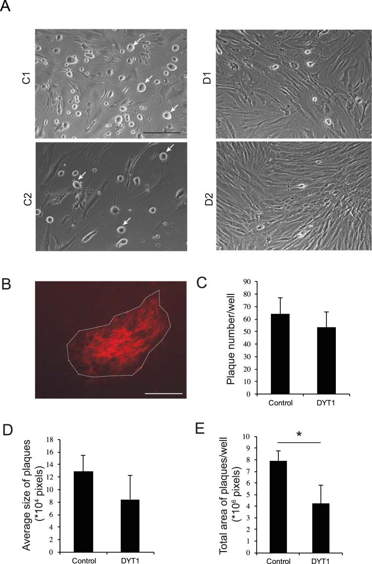
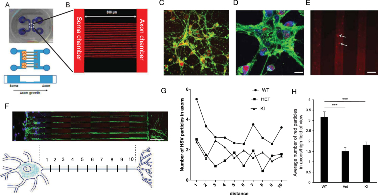
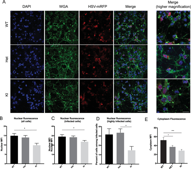
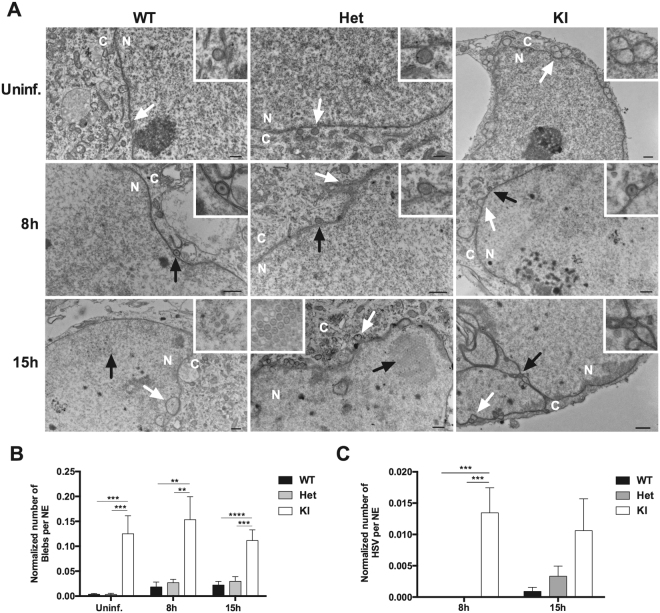
Most cases of early onset torsion dystonia (DYT1) are caused by a 3-base pair deletion in one allele of the TOR1A gene causing loss of a glutamate in torsinA, a luminal protein in the nuclear envelope. This dominantly inherited neurologic disease has reduced penetrance and no other medical manifestations. It has been challenging to understand the neuronal abnormalities as cells and mouse models which are heterozygous (Het) for the mutant allele are quite similar to wild-type (WT) controls. Here we found that patient fibroblasts and mouse neurons Het for this mutation showed significant differences from WT cells in several parameters revealed by infection with herpes simplex virus type 1 (HSV) which replicates in the nucleus and egresses out through the nuclear envelope. Using a red fluorescent protein capsid to monitor HSV infection, patient fibroblasts showed decreased viral plaque formation as compared to controls. Mouse Het neurons had a decrease in cytoplasmic, but not nuclear HSV fluorescence, and reduced numbers of capsids entering axons as compared to infected WT neurons. These findings point to altered dynamics of the nuclear envelope in cells with the patient genotype, which can provide assays to screen for therapeutic agents that can normalize these cells.
大多数早发性扭转痉挛(DYT1)病例是由 TOR1A 基因一个等位基因中的 3 个碱基对缺失引起的,导致核膜内腔蛋白 torsinA 中的谷氨酸缺失。这种显性遗传的神经疾病具有较低的外显率,没有其他医学表现。由于细胞和杂合突变等位基因的小鼠模型与野生型(WT)对照非常相似,因此理解神经元异常一直具有挑战性。在这里,我们发现感染单纯疱疹病毒 1 型(HSV)的患者成纤维细胞和杂合突变的小鼠神经元与 WT 细胞在几个参数上存在显著差异,HSV 在细胞核内复制并通过核膜逸出。使用红色荧光蛋白衣壳来监测 HSV 感染,与对照相比,患者成纤维细胞的病毒斑形成减少。与感染的 WT 神经元相比,杂合突变的小鼠神经元中的细胞质 HSV 荧光减少,但核 HSV 荧光没有减少,进入轴突的衣壳数量减少。这些发现表明患者基因型的细胞核膜动力学发生改变,这可以提供用于筛选可使这些细胞正常化的治疗剂的检测方法。