• 文献检索
  • 文档翻译
  • 深度研究
  • 学术资讯
  • Suppr Zotero 插件Zotero 插件
  • 邀请有礼
  • 套餐&价格
  • 历史记录
应用&插件
Suppr Zotero 插件Zotero 插件浏览器插件Mac 客户端Windows 客户端微信小程序
定价
高级版会员购买积分包购买API积分包
服务
文献检索文档翻译深度研究API 文档MCP 服务
关于我们
关于 Suppr公司介绍联系我们用户协议隐私条款
关注我们

Suppr 超能文献

核心技术专利:CN118964589B侵权必究
粤ICP备2023148730 号-1Suppr @ 2026

文献检索

告别复杂PubMed语法,用中文像聊天一样搜索,搜遍4000万医学文献。AI智能推荐,让科研检索更轻松。

立即免费搜索

文件翻译

保留排版,准确专业,支持PDF/Word/PPT等文件格式,支持 12+语言互译。

免费翻译文档

深度研究

AI帮你快速写综述,25分钟生成高质量综述,智能提取关键信息,辅助科研写作。

立即免费体验

哪些 MRI 特征提示为恶性纯间质子宫肿瘤而非伴有囊性变的子宫肌瘤?

What MRI features suspect malignant pure mesenchymal uterine tumors rather than uterine leiomyoma with cystic degeneration?

机构信息

Department of Radiology, Seoul National University College of Medicine, Seoul, Korea.

Department of Obstetrics and Gynecology, Cancer Research Institute, Seoul National University College of Medicine, Seoul, Korea.

出版信息

J Gynecol Oncol. 2018 May;29(3):e26. doi: 10.3802/jgo.2018.29.e26. Epub 2018 Jan 4.

DOI:10.3802/jgo.2018.29.e26
PMID:29400019
原文链接:https://pmc.ncbi.nlm.nih.gov/articles/PMC5920213/
Abstract

OBJECTIVE

To retrospectively assess conventional magnetic resonance imaging (MRI) features that differentiate malignant pure mesenchymal uterine tumors (MPMUT); endometrial stromal sarcoma (ESS) and leiomyosarcoma (LMS) from uterine leiomyoma with cystic degeneration (ULCD).

METHODS

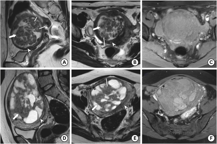
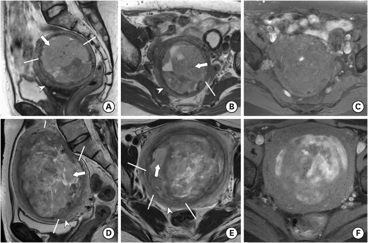
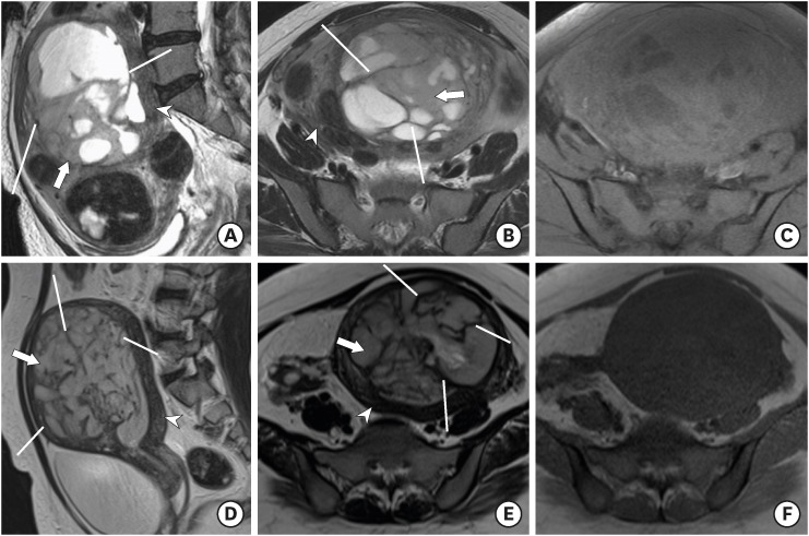
We retrospectively reviewed magnetic resonance (MR) images of 30 patients with ULCD, 18 with ESS, and 15 with LMS, to assess tumor location, margin, T2 signal intensity (SI), speckled appearance, and peripheral band using univariate and multivariate analyses.

RESULTS

ULCD more frequently showed subserosal location (53.3%), well-defined margin (96.7%), and speckled appearance (90.0%) compared with ESS (0%, 33.3%, and 33.3%, respectively) or LMS (20.0%, 33.3%, and 60.0%, respectively). In quantitative T2 SI comparisons, the T2 SI ratio of the main solid tumor portion to gluteus maximus muscle differed significantly among the three groups, with ULCD showing a lower SI ratio (0.62) compared with ESS (2.44) and LMS (1.13). On multivariate analysis, an ill-defined margin (odds ratio [OR]=44.885; p=0.003) and high T2 SI (OR=4.396; p=0.046) were the significant MR differentiators.

CONCLUSION

An ill-defined tumor margin and high T2 SI ratio of the main solid tumor-to-gluteus maximus muscle were useful MRI features in the differentiation of MPMUT from ULCD.

摘要

目的

回顾性评估常规磁共振成像(MRI)特征,以区分恶性单纯间质性子宫肿瘤(MPMUT);子宫内膜间质肉瘤(ESS)和平滑肌肉瘤(LMS)与囊性变性的平滑肌瘤(ULCD)。

方法

我们回顾性分析了 30 例 ULCD、18 例 ESS 和 15 例 LMS 患者的磁共振(MR)图像,以评估肿瘤位置、边缘、T2 信号强度(SI)、斑点外观和外周带,采用单变量和多变量分析。

结果

与 ESS(0%、33.3%和 33.3%)或 LMS(20.0%、33.3%和 60.0%)相比,ULCD 更常表现为子宫浆膜下位置(53.3%)、清晰的边界(96.7%)和斑点外观(90.0%)。在定量 T2 SI 比较中,三组之间主实质肿瘤部分与臀大肌的 T2 SI 比值差异有统计学意义,ULCD 显示较低的 SI 比值(0.62),与 ESS(2.44)和 LMS(1.13)相比。多变量分析显示,边界不清(比值比[OR]=44.885;p=0.003)和 T2 SI 高(OR=4.396;p=0.046)是有意义的 MRI 鉴别特征。

结论

肿瘤边界不清和主实质肿瘤与臀大肌的 T2 SI 比值高是 MPMUT 与 ULCD 鉴别诊断的有用 MRI 特征。

https://cdn.ncbi.nlm.nih.gov/pmc/blobs/99fa/5920213/c36d0ded4116/jgo-29-e26-g003.jpg
https://cdn.ncbi.nlm.nih.gov/pmc/blobs/99fa/5920213/620bc5956b24/jgo-29-e26-g001.jpg
https://cdn.ncbi.nlm.nih.gov/pmc/blobs/99fa/5920213/e5c0ee9f802e/jgo-29-e26-g002.jpg
https://cdn.ncbi.nlm.nih.gov/pmc/blobs/99fa/5920213/c36d0ded4116/jgo-29-e26-g003.jpg
https://cdn.ncbi.nlm.nih.gov/pmc/blobs/99fa/5920213/620bc5956b24/jgo-29-e26-g001.jpg
https://cdn.ncbi.nlm.nih.gov/pmc/blobs/99fa/5920213/e5c0ee9f802e/jgo-29-e26-g002.jpg
https://cdn.ncbi.nlm.nih.gov/pmc/blobs/99fa/5920213/c36d0ded4116/jgo-29-e26-g003.jpg

相似文献

1
What MRI features suspect malignant pure mesenchymal uterine tumors rather than uterine leiomyoma with cystic degeneration?哪些 MRI 特征提示为恶性纯间质子宫肿瘤而非伴有囊性变的子宫肌瘤?
J Gynecol Oncol. 2018 May;29(3):e26. doi: 10.3802/jgo.2018.29.e26. Epub 2018 Jan 4.
2
MRI appearance of mesenchymal tumors of the uterus.子宫间质肿瘤的 MRI 表现。
Eur J Radiol. 2010 Apr;74(1):241-9. doi: 10.1016/j.ejrad.2009.03.005. Epub 2009 Apr 5.
3
Assessment of MR Imaging as a Tool to Differentiate between the Major Histological Types of Uterine Sarcomas.磁共振成像作为鉴别子宫肉瘤主要组织学类型工具的评估
Magn Reson Med Sci. 2015;14(4):295-304. doi: 10.2463/mrms.2014-0023. Epub 2015 Jun 23.
4
Uterine smooth muscle tumours with hyperintense area on T weighted images: differentiation between leiomyosarcomas and leiomyomas.T加权图像上有高信号区的子宫平滑肌肿瘤:平滑肌肉瘤与平滑肌瘤的鉴别
Br J Radiol. 2018 Apr;91(1084):20170767. doi: 10.1259/bjr.20170767. Epub 2018 Jan 18.
5
Multiparametric MR evaluation of uterine leiomyosarcoma and STUMP versus leiomyoma in symptomatic women planned for high frequency focussed ultrasound: accuracy of imaging parameters and interobserver agreement for identification of malignancy.磁共振多参数评价计划行高聚焦超声治疗的有症状女性的子宫平滑肌肉瘤和子宫平滑肌瘤 STUMP 与平滑肌瘤:影像参数的准确性和识别恶性肿瘤的观察者间一致性。
Br J Radiol. 2021 Mar 1;94(1119):20200483. doi: 10.1259/bjr.20200483. Epub 2021 Jan 28.
6
Magnetic resonance imaging features of uterine sarcoma and mimickers.磁共振成像在子宫肉瘤及类似病变中的特征表现。
Abdom Radiol (NY). 2017 Jun;42(6):1762-1772. doi: 10.1007/s00261-017-1076-9.
7
MR imaging findings of unusual leiomyoma and malignant uterine myometrial tumors: what the radiologist should know.磁共振成像对罕见的平滑肌瘤和恶性子宫平滑肌肿瘤的表现:放射科医生应该知道的。
Jpn J Radiol. 2021 Jun;39(6):527-539. doi: 10.1007/s11604-021-01096-7. Epub 2021 Jan 31.
8
MRI screening for uterine leiomyosarcoma.MRI 筛查子宫平滑肌肉瘤。
J Magn Reson Imaging. 2019 Jun;49(7):e282-e294. doi: 10.1002/jmri.26630. Epub 2019 Jan 13.
9
Differentiation of Uterine Leiomyosarcoma from Atypical Leiomyoma: Diagnostic Accuracy of Qualitative MR Imaging Features and Feasibility of Texture Analysis.子宫平滑肌肉瘤与非典型平滑肌瘤的鉴别:定性磁共振成像特征的诊断准确性及纹理分析的可行性
Eur Radiol. 2017 Jul;27(7):2903-2915. doi: 10.1007/s00330-016-4623-9. Epub 2016 Dec 5.
10
The role of multiparametric MRI in differentiating uterine leiomyosarcoma from benign degenerative leiomyoma and leiomyoma variants: a retrospective analysis.多参数 MRI 在鉴别子宫平滑肌肉瘤与良性退行性平滑肌瘤及平滑肌瘤变异型中的作用:一项回顾性分析。
Clin Radiol. 2023 Jan;78(1):47-54. doi: 10.1016/j.crad.2022.08.144. Epub 2022 Oct 8.

引用本文的文献

1
A Rare Case of Low-Grade Endometrial Stromal Sarcoma Invading an Old Leiomyoma.1例罕见的低级别子宫内膜间质肉瘤侵犯陈旧性平滑肌瘤病例
Cureus. 2024 Nov 9;16(11):e73329. doi: 10.7759/cureus.73329. eCollection 2024 Nov.
2
A case of low-grade endometrial stromal sarcoma presented as an intramyometrial mass mimicking uterine leiomyoma on MRI.一例低级别子宫内膜间质肉瘤表现为肌层内肿块,在磁共振成像(MRI)上酷似子宫平滑肌瘤。
BJR Case Rep. 2023 Dec 18;10(1):uaad012. doi: 10.1093/bjrcr/uaad012. eCollection 2024 Jan.
3
Differentiating Uterine Sarcoma From Atypical Leiomyoma on Preoperative Magnetic Resonance Imaging Using Logistic Regression Classifier: Added Value of Diffusion-Weighted Imaging-Based Quantitative Parameters.

本文引用的文献

1
Morcellation and the Incidence of Occult Uterine Malignancy: A Dual-Institution Review.碎宫术与隐匿性子宫恶性肿瘤的发生率:一项双机构回顾研究
Int J Gynecol Cancer. 2016 Jan;26(1):149-55. doi: 10.1097/IGC.0000000000000558.
2
Clinical characteristics and management experience of unexpected uterine sarcoma after myomectomy.子宫肌瘤剔除术后意外子宫肉瘤的临床特征及治疗经验
Int J Gynaecol Obstet. 2015 Aug;130(2):195-9. doi: 10.1016/j.ijgo.2015.01.009. Epub 2015 Mar 24.
3
Assessment of MR Imaging as a Tool to Differentiate between the Major Histological Types of Uterine Sarcomas.
术前磁共振成像中利用逻辑回归分类器鉴别子宫肉瘤与非典型平滑肌瘤:扩散加权成像定量参数的附加价值。
Korean J Radiol. 2024 Jan;25(1):43-54. doi: 10.3348/kjr.2023.0760.
4
MRI Evaluation of Uterine Masses for Risk of Leiomyosarcoma: A Consensus Statement.MRI 评估子宫肿块发生平滑肌肉瘤的风险:共识声明。
Radiology. 2023 Feb;306(2):e211658. doi: 10.1148/radiol.211658. Epub 2022 Oct 4.
5
Advances in the Preoperative Identification of Uterine Sarcoma.子宫肉瘤术前诊断的进展
Cancers (Basel). 2022 Jul 20;14(14):3517. doi: 10.3390/cancers14143517.
6
Uterine fibroid-like tumors: spectrum of MR imaging findings and their differential diagnosis.子宫平滑肌瘤样肿瘤:磁共振成像表现谱及其鉴别诊断
Abdom Radiol (NY). 2022 Jun;47(6):2197-2208. doi: 10.1007/s00261-022-03431-6. Epub 2022 Mar 26.
7
Utility of magnetic resonance imaging for differentiating malignant mesenchymal tumors of the uterus from T2-weighted hyperintense leiomyomas.磁共振成像在鉴别子宫恶性间叶肿瘤与 T2 加权高信号平滑肌瘤中的应用。
Jpn J Radiol. 2022 Apr;40(4):385-395. doi: 10.1007/s11604-021-01217-2. Epub 2021 Nov 9.
8
Differentiation of uterine low-grade endometrial stromal sarcoma from rare leiomyoma variants by magnetic resonance imaging.磁共振成像在子宫低级别子宫内膜间质肉瘤与罕见的平滑肌瘤变异型中的鉴别诊断。
Sci Rep. 2021 Sep 27;11(1):19124. doi: 10.1038/s41598-021-98473-z.
9
The Value of Magnetic Resonance Imaging Histograms in the Preoperative Differential Diagnosis of Endometrial Stromal Sarcoma and Degenerative Hysteromyoma.磁共振成像直方图在子宫内膜间质肉瘤与变性子宫平滑肌瘤术前鉴别诊断中的价值
Front Surg. 2021 Sep 10;8:726067. doi: 10.3389/fsurg.2021.726067. eCollection 2021.
10
Multiparametric MR evaluation of uterine leiomyosarcoma and STUMP versus leiomyoma in symptomatic women planned for high frequency focussed ultrasound: accuracy of imaging parameters and interobserver agreement for identification of malignancy.磁共振多参数评价计划行高聚焦超声治疗的有症状女性的子宫平滑肌肉瘤和子宫平滑肌瘤 STUMP 与平滑肌瘤:影像参数的准确性和识别恶性肿瘤的观察者间一致性。
Br J Radiol. 2021 Mar 1;94(1119):20200483. doi: 10.1259/bjr.20200483. Epub 2021 Jan 28.
磁共振成像作为鉴别子宫肉瘤主要组织学类型工具的评估
Magn Reson Med Sci. 2015;14(4):295-304. doi: 10.2463/mrms.2014-0023. Epub 2015 Jun 23.
4
Uterine sarcomas: clinical presentation and MRI features.子宫肉瘤:临床表现及磁共振成像特征
Diagn Interv Radiol. 2015 Jan-Feb;21(1):4-9. doi: 10.5152/dir.2014.14053.
5
Power morcellators: a review of current practice and assessment of risk.电动组织粉碎器:当前实践的回顾和风险评估。
Am J Obstet Gynecol. 2015 Jan;212(1):18-23. doi: 10.1016/j.ajog.2014.07.046. Epub 2014 Jul 26.
6
Uterine pathology in women undergoing minimally invasive hysterectomy using morcellation.接受使用粉碎术的微创子宫切除术的女性的子宫病理学
JAMA. 2014 Sep 24;312(12):1253-5. doi: 10.1001/jama.2014.9005.
7
Magnetic resonance and diffusion-weighted imaging in categorization of uterine sarcomas: correlation with pathological findings.磁共振成像和扩散加权成像在子宫肉瘤分类中的应用:与病理结果的相关性
Clin Imaging. 2014 Nov-Dec;38(6):836-44. doi: 10.1016/j.clinimag.2014.06.004. Epub 2014 Jun 12.
8
How to differentiate benign from malignant myometrial tumours using MR imaging.如何通过磁共振成像区分良性和恶性子宫肌肿瘤。
Eur Radiol. 2013 Aug;23(8):2306-14. doi: 10.1007/s00330-013-2819-9. Epub 2013 Apr 8.
9
Risk of occult malignancy in morcellated hysterectomy: a case series.经子宫分碎术的隐匿性恶性肿瘤风险:病例系列研究。
Int J Gynecol Pathol. 2011 Sep;30(5):476-83. doi: 10.1097/PGP.0b013e3182107ecf.
10
Endometrial stromal sarcoma located in the myometrium with a low-intensity rim on T2-weighted images: report of three cases and literature review.T2 加权图像显示子宫肌层中存在低信号环的子宫内膜间质肉瘤:三例报告及文献复习。
J Magn Reson Imaging. 2010 Apr;31(4):975-9. doi: 10.1002/jmri.22126.