Eppelheimer Maggie S, Houston James R, Bapuraj Jayapalli R, Labuda Richard, Loth Dorothy M, Braun Audrey M, Allen Natalie J, Heidari Pahlavian Soroush, Biswas Dipankar, Urbizu Aintzane, Martin Bryn A, Maher Cormac O, Allen Philip A, Loth Francis
Department of Biomedical Engineering, Conquer Chiari Research Center, University of Akron, Akron, OH, United States.
Department of Psychology, Conquer Chiari Research Center, University of Akron, Akron, OH, United States.
Front Neuroanat. 2018 Jan 19;12:2. doi: 10.3389/fnana.2018.00002. eCollection 2018.
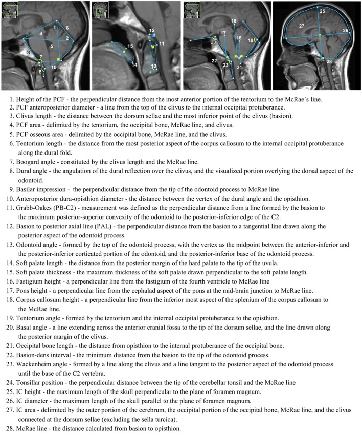
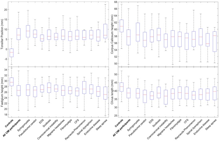
Researchers have sought to better understand Chiari type I malformation (CMI) through morphometric measurements beyond tonsillar position (TP). Soft tissue and bone structures within the brain and craniocervical junction have been shown to be different for CMI patients compared to healthy controls. Yet, several morphological characteristics have not been consistently associated with CMI. CMI is also associated with different prevalent conditions (PCs) such as syringomyelia, pseudotumor, Ehlers-Danlos syndrome (EDS), scoliosis, and craniocervical instability. The goal of this study was two-fold: (1) to identify unique morphological characteristics of PCs, and (2) to better explain inconsistent results from case-control comparisons of CMI. Image, demographic, and PC information was obtained through the , a self-report web-accessed database. Twenty-eight morphometric measurements (MMs) were performed on the cranial MR images of 236 pre-surgery adult female CMI participants and 140 female healthy control participants. Custom software was used to measure 28 structures within the posterior cranial fossa (PCF) compartment, craniocervical junction, oral cavity, and intracranial area on midsagittal MR images for each participant. Morphometric analysis of adult females indicated a smaller McRae line length in CMI participants with syringomyelia compared to those without syringomyelia. TP was reduced in CMI participants with EDS than those without EDS. Basion to posterior axial line was significantly longer in CMI participants with scoliosis compared to those without scoliosis. No additional MMs were found to differ between CMI participants with and without a specific PC. Four morphometric differences were found to be consistently different between CMI participants and healthy controls regardless of PC: larger TP and a smaller clivus length, fastigium, and corpus callosum height in CMI participants. Syringomyelia, EDS, and scoliosis were the only PCs that showed significant morphometric differences between CMI participants. Additionally, four midsagittal MR-based MMs were found to be significantly different between healthy controls and CMI participants regardless of the presence of one or more PCs. This study suggests that the prevalence of comorbid conditions are not strongly related to CMI morphology, and that inconsistent findings in the radiographic literature cannot be explained by varying prevalence of comorbid conditions in CMI study samples.
研究人员试图通过扁桃体位置(TP)以外的形态测量来更好地理解 Chiari I 型畸形(CMI)。与健康对照相比,CMI 患者脑内及颅颈交界处的软组织和骨骼结构已被证明存在差异。然而,一些形态学特征与 CMI 的关联并不一致。CMI 还与不同的常见病症(PCs)相关,如脊髓空洞症、假瘤、埃勒斯-当洛综合征(EDS)、脊柱侧弯和颅颈不稳定。本研究的目标有两个:(1)识别常见病症的独特形态学特征,(2)更好地解释 CMI 病例对照比较中不一致的结果。图像、人口统计学和常见病症信息通过一个可通过网络自助访问的数据库获取。对 236 名术前成年女性 CMI 参与者和 140 名女性健康对照参与者的头颅磁共振图像进行了 28 项形态测量(MMs)。使用定制软件在每位参与者的矢状面磁共振图像上测量后颅窝(PCF)腔、颅颈交界处、口腔和颅内区域内的 28 个结构。成年女性的形态测量分析表明,与无脊髓空洞症的 CMI 参与者相比,有脊髓空洞症的 CMI 参与者的麦克雷线长度更小。与无 EDS 的 CMI 参与者相比,有 EDS 的 CMI 参与者的 TP 降低。与无脊柱侧弯的 CMI 参与者相比,有脊柱侧弯的 CMI 参与者的颅底至后轴线明显更长。在有无特定常见病症的 CMI 参与者之间未发现其他形态测量有差异。无论常见病症如何,在 CMI 参与者和健康对照之间发现有四个形态测量差异始终存在:CMI 参与者的 TP 更大,斜坡长度、小脑蚓部顶点和胼胝体高度更小。脊髓空洞症、EDS 和脊柱侧弯是仅有的在 CMI 参与者之间显示出显著形态测量差异的常见病症。此外,无论是否存在一种或多种常见病症,在健康对照和 CMI 参与者之间发现基于矢状面磁共振图像的四个形态测量有显著差异。本研究表明,合并症的患病率与 CMI 形态学没有密切关系,并且放射学文献中不一致的发现不能用 CMI 研究样本中合并症患病率的差异来解释。