University of Houston, College of Optometry, 4901 Calhoun Road, Houston, TX 77204, USA.
University of Houston, College of Optometry, 4901 Calhoun Road, Houston, TX 77204, USA.
Exp Eye Res. 2018 Apr;169:79-90. doi: 10.1016/j.exer.2018.01.025. Epub 2018 Feb 1.
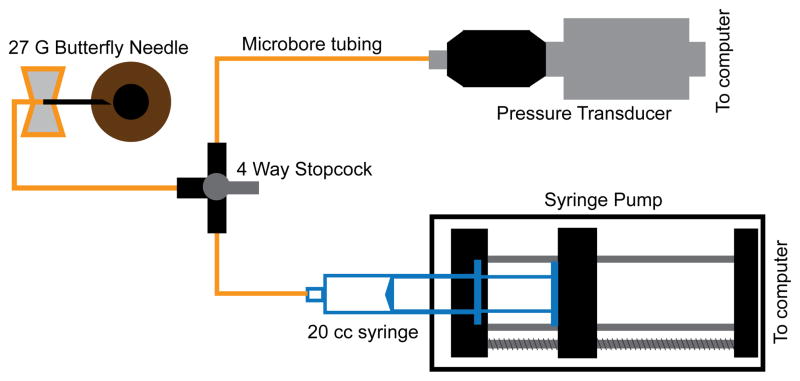
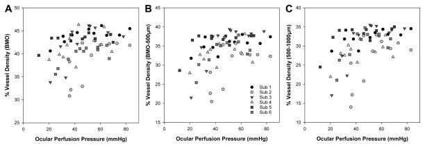
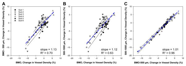
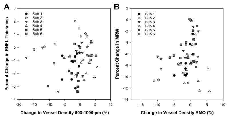
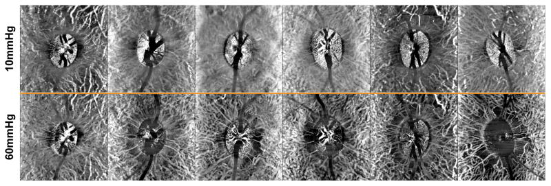
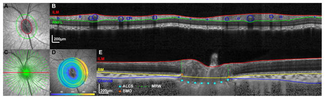
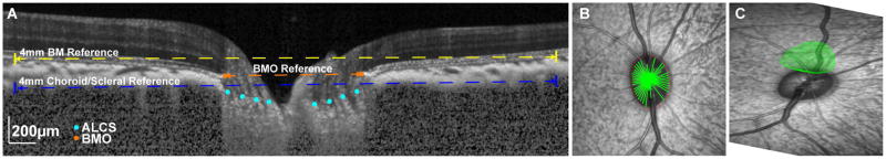
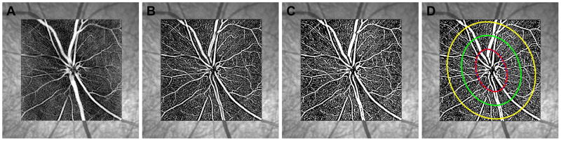
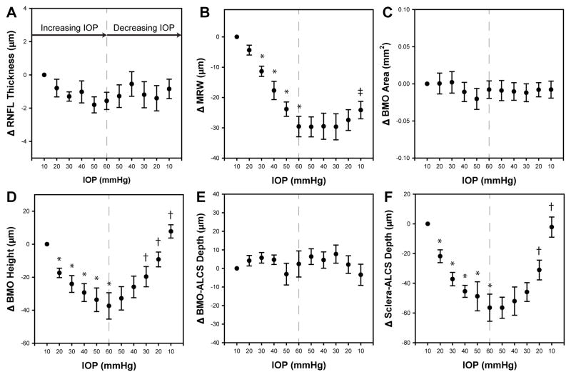
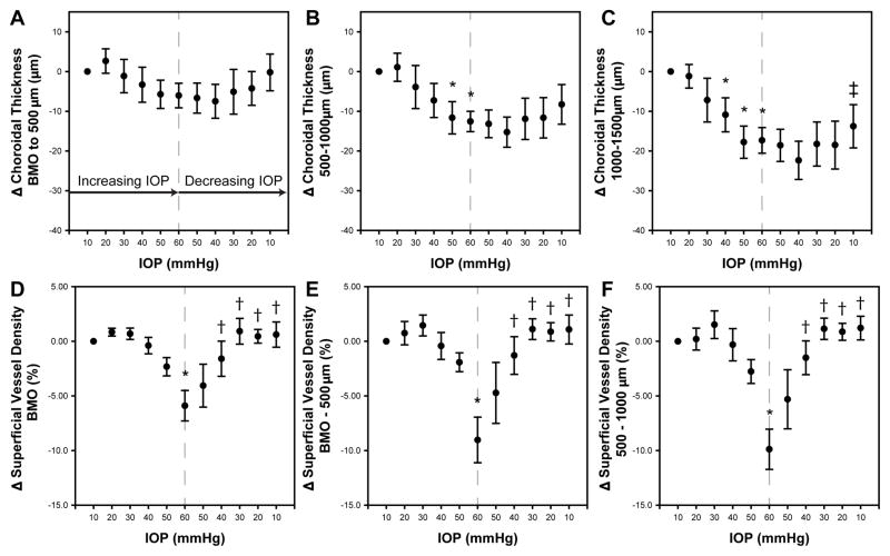
Intraocular pressure (IOP) is an important risk factor for glaucoma, and the response of the ONH and surrounding tissues to elevated IOP are often investigated to better understand pathophysiology. In vivo structure including that of the optic nerve head (ONH) and surrounding tissue of the eye are often assessed using optical coherence tomography (OCT). With advances in OCT technology, both large vessels and capillaries can be imaged non-invasively (OCT Angiography). Because a significant portion of retinal thickness is comprised of vasculature, the purpose of the current study was to investigate OCT structural and vascular changes in healthy non-human primate eyes with systematic graded increases and decreases in IOP. Six healthy animals with no previous experimental intervention were used. The pressure in the anterior chamber was adjusted from 10 mmHg to 60 mmHg and back to 10 mmHg in 10 mmHg steps every 10 min. Using optical coherence tomography (OCT), retinal nerve fiber layer (RNFL) thickness, minimum rim width (MRW), Bruch's membrane opening (BMO) size and relative height, anterior lamina cribrosa surface (ALCS) depth, choroidal thickness, and angiography (OCTA) were quantified. With IOP challenge there were significant changes in all morphological measures quantified (p < 0.01) other than BMO size (p = 0.30) and RNFL thickness (p = 0.29). Specifically, the position of the BMO was sensitive to both an increase and decease in IOP. The inner retinal capillary density gradually decreased with increasing IOP, reaching statistical significance when pressure exceeded 50 mmHg, but returned when IOP was reduced. The average choroidal thickness around the ONH decreased for elliptical annuli 500-1000 μm and 1000-1500 μm, from the BMO, with increasing IOP (p < 0.01). For the 1000-1500 μm annulus, choroid thickness did not return to baseline with IOP reduction. Similarly, the MRW decreased with increase in IOP, but with pressure reduction did not return, and at the final 10 mmHg time point was thinner than at baseline (p < 0.01). The results from this experiment illustrate differences in ONH neural rim tissue, RNFL and vessel density changes with acute IOP challenge. Overall, vessel collapse could not completely account for changes in RNFL or ONH MRW thickness. The study supports the hypothesis neural rim compression may be an important part of IOP-induced damage.
眼压(IOP)是青光眼的一个重要危险因素,因此人们经常研究视盘(ONH)及其周围组织对眼压升高的反应,以更好地了解其病理生理学机制。通常使用光学相干断层扫描(OCT)来评估活体结构,包括视神经头(ONH)和眼部周围组织。随着 OCT 技术的进步,现在可以无创地成像大血管和毛细血管(OCT 血管造影)。由于视网膜厚度的很大一部分由血管组成,因此本研究的目的是在健康的非人类灵长类动物中,通过系统地逐步升高和降低眼压,来研究 OCT 结构和血管的变化。使用了 6 只未进行过先前实验干预的健康动物。每隔 10 分钟,将前房压力从 10mmHg 升高到 60mmHg,然后再升高到 10mmHg。使用光学相干断层扫描(OCT),测量视网膜神经纤维层(RNFL)厚度、最小 rim 宽度(MRW)、Bruch 膜开口(BMO)大小和相对高度、前 lamina cribrosa 表面(ALCS)深度、脉络膜厚度和血管造影(OCTA)。随着眼压的升高,所有形态学测量值都有显著变化(p<0.01),除了 BMO 大小(p=0.30)和 RNFL 厚度(p=0.29)。具体来说,BMO 的位置对眼压的升高和降低都很敏感。随着眼压的升高,内视网膜毛细血管密度逐渐降低,当压力超过 50mmHg 时达到统计学意义,但当眼压降低时又恢复。从 BMO 开始,ONH 周围的脉络膜厚度在 500-1000μm 和 1000-1500μm 的椭圆形环中逐渐减小,随着眼压的升高(p<0.01)。对于 1000-1500μm 的环,当眼压降低时,脉络膜厚度并未恢复到基线水平。同样,MRW 随着眼压的升高而减小,但随着压力的降低并未恢复,并且在最后一个 10mmHg 时间点比基线时更薄(p<0.01)。该实验的结果表明,急性眼压升高会导致 ONH 神经边缘组织、RNFL 和血管密度发生变化。总的来说,血管塌陷不能完全解释 RNFL 或 ONH MRW 厚度的变化。该研究支持神经边缘组织受压可能是眼压引起损伤的一个重要因素的假说。