• 文献检索
  • 文档翻译
  • 深度研究
  • 学术资讯
  • Suppr Zotero 插件Zotero 插件
  • 邀请有礼
  • 套餐&价格
  • 历史记录
应用&插件
Suppr Zotero 插件Zotero 插件浏览器插件Mac 客户端Windows 客户端微信小程序
定价
高级版会员购买积分包购买API积分包
服务
文献检索文档翻译深度研究API 文档MCP 服务
关于我们
关于 Suppr公司介绍联系我们用户协议隐私条款
关注我们

Suppr 超能文献

核心技术专利:CN118964589B侵权必究
粤ICP备2023148730 号-1Suppr @ 2026

文献检索

告别复杂PubMed语法,用中文像聊天一样搜索,搜遍4000万医学文献。AI智能推荐,让科研检索更轻松。

立即免费搜索

文件翻译

保留排版,准确专业,支持PDF/Word/PPT等文件格式,支持 12+语言互译。

免费翻译文档

深度研究

AI帮你快速写综述,25分钟生成高质量综述,智能提取关键信息,辅助科研写作。

立即免费体验

神经视网膜边缘对瞬态眼压挑战的反应可预测实验性青光眼中视网膜神经节细胞损失的程度。

Neuroretinal Rim Response to Transient Intraocular Pressure Challenge Predicts the Extent of Retinal Ganglion Cell Loss in Experimental Glaucoma.

机构信息

University of Houston College of Optometry, Houston, Texas, United States.

出版信息

Invest Ophthalmol Vis Sci. 2023 May 1;64(5):30. doi: 10.1167/iovs.64.5.30.

DOI:10.1167/iovs.64.5.30
PMID:37256608
原文链接:https://pmc.ncbi.nlm.nih.gov/articles/PMC10233313/
Abstract

PURPOSE

To determine if the optic nerve head (ONH) response to transient elevated intraocular pressure (IOP) can predict the extent of neural loss in the nonhuman primate experimental glaucoma model.

METHODS

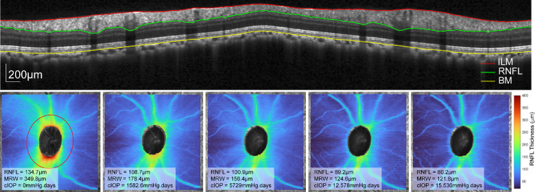
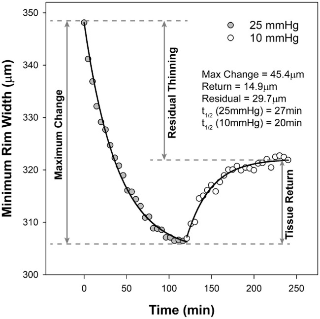
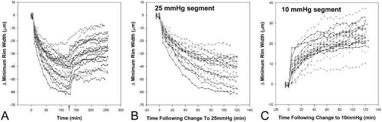
The anterior chamber pressure of 21 healthy animals (5.4 ± 1.2 years, 8 female) was adjusted to 25 mm Hg for two hours followed by 10 mm Hg for an additional two hours. For the duration of IOP challenge the ONH was imaged using radial optical coherence tomography (OCT) scans at five-minute intervals. Afterward, a randomized sample of 14 of these subjects had unilateral experimental glaucoma induced and were monitored with OCT imaging, tonometry, and ocular biometry at two-week intervals.

RESULTS

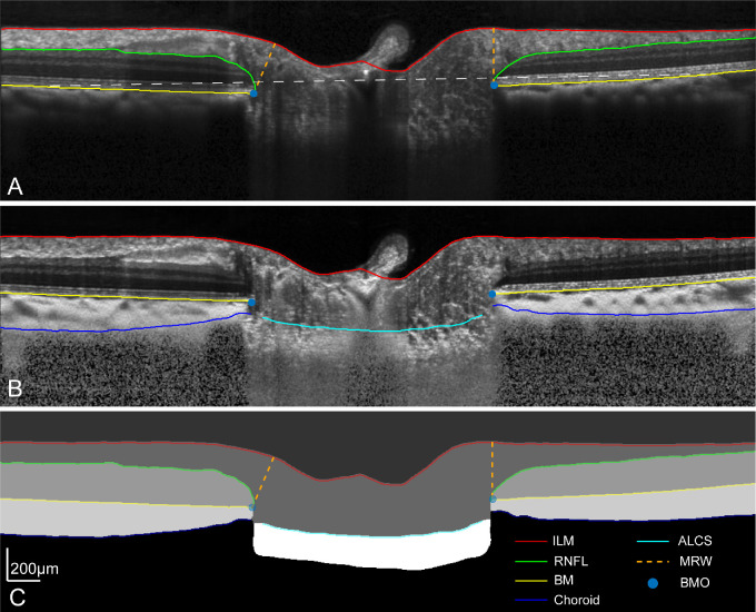
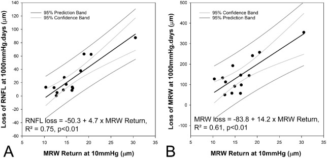
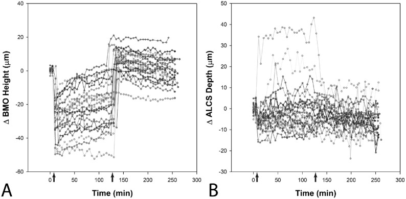
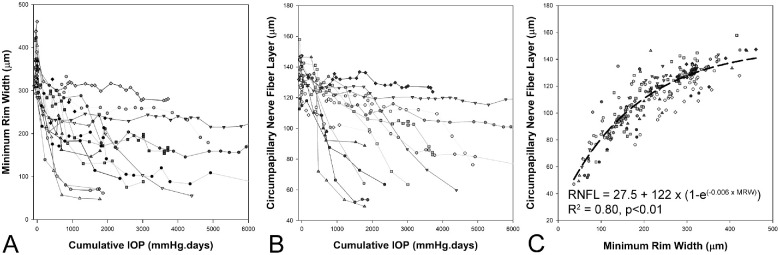
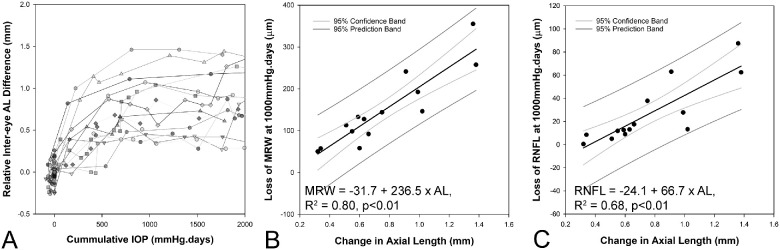
With pressure challenge, the maximum decrease in ONH minimum rim width (MRW) was 40 ± 10.5 µm at 25 mm Hg and was correlated with the precannulation MRW, Bruch's membrane opening (BMO) position, and the anterior lamina cribrosa surface position (P = 0.01). The maximum return of MRW at 10 mm Hg was 16.1 ± 5.0 µm and was not associated with any precannulation ONH feature (P = 0.24). However, healthy eyes with greater thickness return at 10 mm Hg had greater loss of MRW and retinal nerve fiber layer (RNFL) at a cumulative IOP of 1000 mm Hg · days after induction of experimental glaucoma. In addition, MRW and RNFL thinning was correlated with an increase in axial length (P < 0.01).

CONCLUSION

This study's findings suggest that the ONH's response to transient changes in IOP are associated with features of the ONH and surrounding tissues. The neural rim properties at baseline and the extent of axial elongation are associated with the severity of glaucomatous loss in the nonhuman primate model.

摘要

目的

确定视神经头(ONH)对短暂性眼内压升高(IOP)的反应是否可以预测非人灵长类实验性青光眼模型中的神经损失程度。

方法

21 只健康动物(5.4 ± 1.2 岁,8 只雌性)的前房压力调整为 25mmHg 持续两小时,然后再调整为 10mmHg 持续两小时。在 IOP 挑战期间,使用径向光相干断层扫描(OCT)以五分钟的间隔对 ONH 进行成像。之后,对其中 14 只随机选择的动物进行单侧实验性青光眼诱导,并通过 OCT 成像、眼压测量和眼部生物测量在两周的间隔进行监测。

结果

在压力挑战期间,ONH 最小 rim 宽度(MRW)的最大下降幅度为 25mmHg 时的 40 ± 10.5µm,与预插管时的 MRW、Bruch 膜开口(BMO)位置和前 lamina cribrosa 表面位置相关(P = 0.01)。在 10mmHg 时 MRW 的最大恢复幅度为 16.1 ± 5.0µm,与任何预插管 ONH 特征均无关(P = 0.24)。然而,在诱导实验性青光眼后累积 IOP 为 1000mmHg·天,10mmHg 时具有更大厚度恢复的健康眼睛,其 MRW 和视网膜神经纤维层(RNFL)的损失更大。此外,MRW 和 RNFL 变薄与眼轴长度的增加相关(P < 0.01)。

结论

本研究的结果表明,ONH 对 IOP 短暂变化的反应与 ONH 和周围组织的特征相关。基线时神经 rim 特性和轴向伸长的程度与非人灵长类模型中青光眼损失的严重程度相关。

https://cdn.ncbi.nlm.nih.gov/pmc/blobs/fc22/10233313/b40db580ff99/iovs-64-5-30-f008.jpg
https://cdn.ncbi.nlm.nih.gov/pmc/blobs/fc22/10233313/12b73d2acb3a/iovs-64-5-30-f001.jpg
https://cdn.ncbi.nlm.nih.gov/pmc/blobs/fc22/10233313/0b26d60528ac/iovs-64-5-30-f002.jpg
https://cdn.ncbi.nlm.nih.gov/pmc/blobs/fc22/10233313/7bc2764f3062/iovs-64-5-30-f003.jpg
https://cdn.ncbi.nlm.nih.gov/pmc/blobs/fc22/10233313/816f34cb2468/iovs-64-5-30-f004.jpg
https://cdn.ncbi.nlm.nih.gov/pmc/blobs/fc22/10233313/3cfed52d8a18/iovs-64-5-30-f005.jpg
https://cdn.ncbi.nlm.nih.gov/pmc/blobs/fc22/10233313/31754100e891/iovs-64-5-30-f006.jpg
https://cdn.ncbi.nlm.nih.gov/pmc/blobs/fc22/10233313/48ef73ab97e1/iovs-64-5-30-f007.jpg
https://cdn.ncbi.nlm.nih.gov/pmc/blobs/fc22/10233313/b40db580ff99/iovs-64-5-30-f008.jpg

相似文献

1
Neuroretinal Rim Response to Transient Intraocular Pressure Challenge Predicts the Extent of Retinal Ganglion Cell Loss in Experimental Glaucoma.神经视网膜边缘对瞬态眼压挑战的反应可预测实验性青光眼中视网膜神经节细胞损失的程度。
Invest Ophthalmol Vis Sci. 2023 May 1;64(5):30. doi: 10.1167/iovs.64.5.30.
2
Neuroretinal rim response to transient changes in intraocular pressure in healthy non-human primate eyes.健康非人类灵长类动物眼内压瞬态变化的神经视网膜边缘反应。
Exp Eye Res. 2020 Apr;193:107978. doi: 10.1016/j.exer.2020.107978. Epub 2020 Feb 17.
3
The effects of graded intraocular pressure challenge on the optic nerve head.眼压递增刺激对视神经乳头的影响。
Exp Eye Res. 2018 Apr;169:79-90. doi: 10.1016/j.exer.2018.01.025. Epub 2018 Feb 1.
4
Evaluation of two-dimensional Bruch's membrane opening minimum rim area for glaucoma diagnostics in a large patient cohort.大样本患者群体中二维 Bruch 膜开口最小边缘区域在青光眼诊断中的评估。
Acta Ophthalmol. 2019 Feb;97(1):60-67. doi: 10.1111/aos.13698. Epub 2018 Mar 24.
5
Effect of Trabeculectomy on OCT Measurements of the Optic Nerve Head Neuroretinal Rim Tissue.小梁切除术对 OCT 测量视神经头神经视网膜边缘组织的影响。
Ophthalmol Glaucoma. 2020 Jan-Feb;3(1):32-39. doi: 10.1016/j.ogla.2019.09.003. Epub 2019 Oct 4.
6
Impact of ab-interno trabeculectomy on Bruch's membrane opening-based morphometry of the optic nerve head for glaucoma progression analysis.内路小梁切除术对基于布鲁赫膜开口的视神经乳头形态测量在青光眼病情进展分析中的影响。
Graefes Arch Clin Exp Ophthalmol. 2019 Feb;257(2):339-347. doi: 10.1007/s00417-018-4187-2. Epub 2018 Nov 28.
7
Novel Bruch's Membrane Opening Minimum Rim Area Equalizes Disc Size Dependency and Offers High Diagnostic Power for Glaucoma.新型布鲁赫膜开口最小边缘面积可平衡视盘大小依赖性并为青光眼提供高诊断效能。
Invest Ophthalmol Vis Sci. 2016 Dec 1;57(15):6596-6603. doi: 10.1167/iovs.16-20561.
8
Comparing optical coherence tomography radial and cube scan patterns for measuring Bruch's membrane opening minimum rim width (BMO-MRW) in glaucoma and healthy eyes: cross-sectional and longitudinal analysis.比较光学相干断层扫描径向和立方扫描模式测量青光眼和正常眼中的 Bruch 膜开口最小边缘宽度(BMO-MRW):横断面和纵向分析。
Br J Ophthalmol. 2018 Mar;102(3):344-351. doi: 10.1136/bjophthalmol-2016-310111. Epub 2017 Aug 3.
9
Effect of acute intraocular pressure elevation on the minimum rim width in normal, ocular hypertensive and glaucoma eyes.急性眼压升高对正常眼、高眼压眼和青光眼眼最小视盘边缘宽度的影响。
Br J Ophthalmol. 2018 Jan;102(1):131-135. doi: 10.1136/bjophthalmol-2017-310232. Epub 2017 May 10.
10
Structure-function relationships with spectral-domain optical coherence tomography retinal nerve fiber layer and optic nerve head measurements.光谱域光学相干断层扫描视网膜神经纤维层和视神经乳头测量的结构-功能关系
Invest Ophthalmol Vis Sci. 2014 May 2;55(5):2953-62. doi: 10.1167/iovs.13-13482.

引用本文的文献

1
Optic Nerve Head Morphology and Macula Ganglion Cell Inner Plexiform Layer Thickness in Axially Anisometropic Rhesus Monkeys.轴向屈光参差恒河猴的视神经头形态和黄斑神经节细胞内丛状层厚度。
Invest Ophthalmol Vis Sci. 2024 Aug 1;65(10):44. doi: 10.1167/iovs.65.10.44.
2
Retinal Ganglion Cell Content Underlying Standard Automated Perimetry Size I to V Visual Sensitivities in the Non-Human Primate Experimental Glaucoma Model.在非人类灵长类实验性青光眼模型中,标准自动视野计大小 I 至 V 视觉敏感度所对应的视网膜神经节细胞含量。
Invest Ophthalmol Vis Sci. 2024 Jul 1;65(8):22. doi: 10.1167/iovs.65.8.22.

本文引用的文献

1
Elevated IOP Alters the Material Properties of Sclera and Lamina Cribrosa in Monkeys.眼压升高改变了猴子巩膜和筛板的材料特性。
Dis Markers. 2022 Aug 23;2022:5038847. doi: 10.1155/2022/5038847. eCollection 2022.
2
Widefield OCT Imaging for Quantifying Inner Retinal Thickness in the Nonhuman Primate.宽视野 OCT 成像在非人类灵长类动物中定量测量内视网膜厚度。
Transl Vis Sci Technol. 2022 Aug 1;11(8):12. doi: 10.1167/tvst.11.8.12.
3
Intraoperative Mounted Optical Coherence Tomography Findings Following Reversal of Optic Nerve Head Cupping in Childhood Glaucoma.
儿童青光眼视神经乳头杯反转术后术中安装的光学相干断层扫描结果。
Am J Ophthalmol. 2022 Nov;243:109-117. doi: 10.1016/j.ajo.2022.08.003. Epub 2022 Aug 8.
4
Short-term changes in Bruch's membrane opening-based morphometrics during the first week after trabeculectomy.在小梁切除术后的第一周内,基于 Bruch 膜开口的形态计量学的短期变化。
Graefes Arch Clin Exp Ophthalmol. 2022 Oct;260(10):3321-3329. doi: 10.1007/s00417-022-05644-3. Epub 2022 Apr 8.
5
The Relationship Between Macula Retinal Ganglion Cell Density and Visual Function in the Nonhuman Primate.灵长类动物的黄斑视网膜神经节细胞密度与视觉功能的关系。
Invest Ophthalmol Vis Sci. 2021 Jan 4;62(1):5. doi: 10.1167/iovs.62.1.5.
6
Neuroretinal rim response to transient changes in intraocular pressure in healthy non-human primate eyes.健康非人类灵长类动物眼内压瞬态变化的神经视网膜边缘反应。
Exp Eye Res. 2020 Apr;193:107978. doi: 10.1016/j.exer.2020.107978. Epub 2020 Feb 17.
7
The Relationship Between Scleral Strain Change and Differential Cumulative Intraocular Pressure Exposure in the Nonhuman Primate Chronic Ocular Hypertension Model.在非人类灵长类慢性高眼压模型中巩膜应变变化与差异累积眼内压暴露的关系。
Invest Ophthalmol Vis Sci. 2019 Oct 1;60(13):4141-4150. doi: 10.1167/iovs.19-27060.
8
Mechanical Deformation of Human Optic Nerve Head and Peripapillary Tissue in Response to Acute IOP Elevation.人眼视神经头和视盘周围组织对急性眼压升高的机械变形。
Invest Ophthalmol Vis Sci. 2019 Mar 1;60(4):913-920. doi: 10.1167/iovs.18-26071.
9
Pulling and Tugging on the Retina: Mechanical Impact of Glaucoma Beyond the Optic Nerve Head.牵拉视网膜:视神经头以外青光眼的机械性影响。
Invest Ophthalmol Vis Sci. 2019 Jan 2;60(1):26-35. doi: 10.1167/iovs.18-25837.
10
In vivo optic nerve head mechanical response to intraocular and cerebrospinal fluid pressure: imaging protocol and quantification method.活体眼内压和脑脊液压力对视盘机械反应的研究:成像方案和定量方法。
Sci Rep. 2018 Aug 23;8(1):12639. doi: 10.1038/s41598-018-31052-x.