• 文献检索
  • 文档翻译
  • 深度研究
  • 学术资讯
  • Suppr Zotero 插件Zotero 插件
  • 邀请有礼
  • 套餐&价格
  • 历史记录
应用&插件
Suppr Zotero 插件Zotero 插件浏览器插件Mac 客户端Windows 客户端微信小程序
定价
高级版会员购买积分包购买API积分包
服务
文献检索文档翻译深度研究API 文档MCP 服务
关于我们
关于 Suppr公司介绍联系我们用户协议隐私条款
关注我们

Suppr 超能文献

核心技术专利:CN118964589B侵权必究
粤ICP备2023148730 号-1Suppr @ 2026

文献检索

告别复杂PubMed语法,用中文像聊天一样搜索,搜遍4000万医学文献。AI智能推荐,让科研检索更轻松。

立即免费搜索

文件翻译

保留排版,准确专业,支持PDF/Word/PPT等文件格式,支持 12+语言互译。

免费翻译文档

深度研究

AI帮你快速写综述,25分钟生成高质量综述,智能提取关键信息,辅助科研写作。

立即免费体验

远红外延时热成像技术对烧伤深度的早期评估

Early Assessment of Burn Depth with Far Infrared Time-Lapse Thermography.

机构信息

Department of Surgery, University of South Alabama School of Medicine, Mobile, AL; Department of Molecular and Cellular Pharmacology, University of South Alabama School of Medicine, Mobile, AL; Center for Lung Biology, University of South Alabama School of Medicine, Mobile, AL.

Department of Surgery, University of South Alabama School of Medicine, Mobile, AL.

出版信息

J Am Coll Surg. 2018 Apr;226(4):687-693. doi: 10.1016/j.jamcollsurg.2017.12.051. Epub 2018 Jan 31.

DOI:10.1016/j.jamcollsurg.2017.12.051
PMID:29409904
原文链接:https://pmc.ncbi.nlm.nih.gov/articles/PMC5880281/
Abstract

BACKGROUND

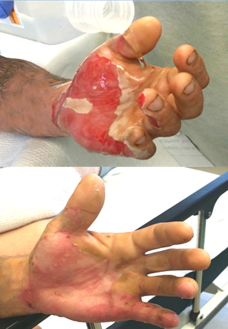
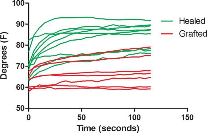
Diagnosing the extremes of superficial burns and full-thickness burns is straightforward. It is in the middle ground of partial-thickness burns where the diagnostic difficulties emerge; it can take up to 3 to 5 days for signs of healing to appear. We hypothesize that cooling partial-thickness burns and tracking the rate of rewarming will immediately reflect the condition of the burn: shallow partial-thickness burns that retain cell health and blood flow will rewarm rapidly, and deeper burns with damaged microvessels will rewarm slowly.

STUDY DESIGN

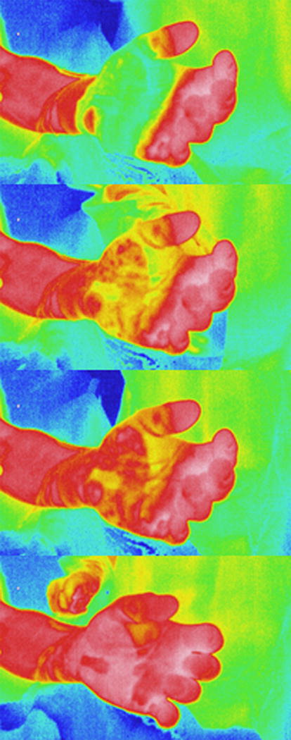
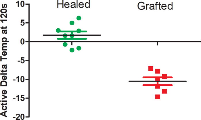
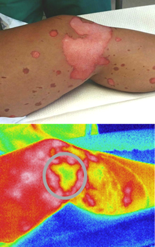
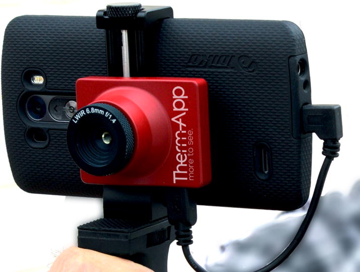
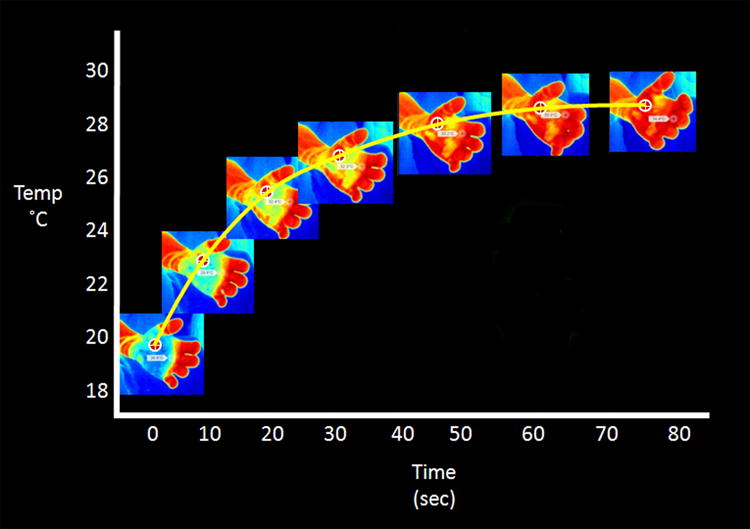
We enrolled 16 patients with isolated, partial-thickness burns on their extremities who were diagnosed as indeterminate by our burn surgeon. Within 24 hours after presentation, room-temperature saline was poured over the burn as a cooling challenge. An infrared camera that was sensitive to body temperature produced false-color images showing pixel-by-pixel temperatures. A time-lapse recording from the infrared camera images taken as the burn rewarmed produced a time-temperature curve that reflected the kinetics of rewarming. The outcomes variable was whether or not the patient received a skin graft, which was determined 72 hours after presentation.

RESULTS

The method correctly predicted whether or not the patient required a skin graft.

CONCLUSIONS

Here we report a new technique that permits determination of wound viability much earlier than clinical examination. Due to the simplicity of the method, non-experts can successfully perform the technique on the first day of the burn and make the correct diagnosis and decision to graft or not to graft.

摘要

背景

诊断表浅烧伤和全层烧伤很简单。在部分厚度烧伤的中间地带出现了诊断困难;直到出现愈合迹象可能需要 3 到 5 天。我们假设冷却部分厚度烧伤并跟踪复温速度将立即反映烧伤的状况:保持细胞健康和血流的浅部分层烧伤会迅速复温,而血管受损的更深烧伤则会缓慢复温。

研究设计

我们招募了 16 名患有孤立性肢体部分厚度烧伤的患者,这些患者被我们的烧伤外科医生诊断为不确定。在出现后 24 小时内,室温生理盐水倒在烧伤处作为冷却挑战。对体温敏感的红外摄像机产生假彩色图像,显示像素级别的温度。从红外摄像机拍摄的烧伤复温的延时记录产生了一个时间-温度曲线,反映了复温的动力学。结局变量是患者是否需要植皮,这是在出现后 72 小时确定的。

结果

该方法正确预测了患者是否需要植皮。

结论

在这里,我们报告了一种新的技术,可以比临床检查更早地确定伤口的存活能力。由于该方法简单,非专家可以在烧伤的第一天成功地进行该技术,并做出正确的诊断和是否植皮的决定。

https://cdn.ncbi.nlm.nih.gov/pmc/blobs/ed71/5880281/6205c8c645e1/nihms944336f10.jpg
https://cdn.ncbi.nlm.nih.gov/pmc/blobs/ed71/5880281/2a7d393ea2c0/nihms944336f1.jpg
https://cdn.ncbi.nlm.nih.gov/pmc/blobs/ed71/5880281/761fe1b7ae19/nihms944336f2.jpg
https://cdn.ncbi.nlm.nih.gov/pmc/blobs/ed71/5880281/54ff6791fe13/nihms944336f3.jpg
https://cdn.ncbi.nlm.nih.gov/pmc/blobs/ed71/5880281/e34dfbf6acf4/nihms944336f4.jpg
https://cdn.ncbi.nlm.nih.gov/pmc/blobs/ed71/5880281/7515db9d41a3/nihms944336f5.jpg
https://cdn.ncbi.nlm.nih.gov/pmc/blobs/ed71/5880281/966eff8c3031/nihms944336f6.jpg
https://cdn.ncbi.nlm.nih.gov/pmc/blobs/ed71/5880281/a6b4d19fc837/nihms944336f7.jpg
https://cdn.ncbi.nlm.nih.gov/pmc/blobs/ed71/5880281/98f963162266/nihms944336f8.jpg
https://cdn.ncbi.nlm.nih.gov/pmc/blobs/ed71/5880281/7f28f0b51b03/nihms944336f9.jpg
https://cdn.ncbi.nlm.nih.gov/pmc/blobs/ed71/5880281/6205c8c645e1/nihms944336f10.jpg

相似文献

1
Early Assessment of Burn Depth with Far Infrared Time-Lapse Thermography.远红外延时热成像技术对烧伤深度的早期评估
J Am Coll Surg. 2018 Apr;226(4):687-693. doi: 10.1016/j.jamcollsurg.2017.12.051. Epub 2018 Jan 31.
2
Forward-looking infrared imaging predicts ultimate burn depth in a porcine vertical injury progression model.前瞻性红外成像可预测猪垂直损伤进展模型中的最终烧伤深度。
Burns. 2016 Mar;42(2):397-404. doi: 10.1016/j.burns.2015.07.006. Epub 2016 Jan 7.
3
A pilot evaluation study of high resolution digital thermal imaging in the assessment of burn depth.高分辨率数字热成像在评估烧伤深度中的初步评估研究。
Burns. 2013 Feb;39(1):76-81. doi: 10.1016/j.burns.2012.03.014. Epub 2012 May 29.
4
Use of FLIR ONE Smartphone Thermography in Burn Wound Assessment.FLIR ONE智能手机热成像技术在烧伤创面评估中的应用
Ann Plast Surg. 2018 Apr;80(4 Suppl 4):S236-S238. doi: 10.1097/SAP.0000000000001363.
5
Infrared Thermal Imaging Has the Potential to Reduce Unnecessary Surgery and Delays to Necessary Surgery in Burn Patients.红外热成像技术有潜力减少烧伤患者不必要的手术,并减少必要手术的延迟。
J Burn Care Res. 2016 Nov/Dec;37(6):350-355. doi: 10.1097/BCR.0000000000000330.
6
Timing of the thermographic assessment of burns.烧伤热成像评估的时机。
Burns. 1996 Feb;22(1):26-8. doi: 10.1016/0305-4179(95)00076-3.
7
Examination of the Early Diagnostic Applicability of Active Dynamic Thermography for Burn Wound Depth Assessment and Concept Analysis.主动动态热成像技术在烧伤创面深度评估中的早期诊断适用性研究及概念分析
J Burn Care Res. 2015 Nov-Dec;36(6):626-35. doi: 10.1097/BCR.0000000000000187.
8
A comparison of non-invasive imaging modalities: Infrared thermography, spectrophotometric intracutaneous analysis and laser Doppler imaging for the assessment of adult burns.非侵入性成像方式的比较:用于评估成人烧伤的红外热成像、分光光度法皮内分析和激光多普勒成像
Burns. 2015 Dec;41(8):1695-1707. doi: 10.1016/j.burns.2015.06.023. Epub 2015 Sep 28.
9
Classification of burn injuries using near-infrared spectroscopy.使用近红外光谱法对烧伤进行分类。
J Biomed Opt. 2006 Sep-Oct;11(5):054002. doi: 10.1117/1.2362722.
10
Evaporative Water Loss in Superficial to Full Thickness Burns.浅度至深度烧伤的蒸发失水量
Ann Plast Surg. 2016 Oct;77(4):401-5. doi: 10.1097/SAP.0000000000000845.

引用本文的文献

1
A Comprehensive Scoping Review on the Use of Point-Of-Care Infrared Thermography Devices for Assessing Various Wound Types.关于使用即时护理红外热成像设备评估各种伤口类型的综合范围综述。
Int Wound J. 2025 Aug;22(8):e70741. doi: 10.1111/iwj.70741.
2
Thermal Imaging as a Method to Indirectly Assess Peripheral Vascular Integrity and Tissue Viability in Veterinary Medicine: Animal Models and Clinical Applications.热成像作为间接评估兽医学中周围血管完整性和组织活力的方法:动物模型与临床应用
Animals (Basel). 2023 Dec 31;14(1):142. doi: 10.3390/ani14010142.
3
The Role of ICG Angiography in Decision Making About Skin-Sparing in Pediatric Acute Trauma.

本文引用的文献

1
Noninvasive Techniques for the Determination of Burn Severity in Real Time.实时测定烧伤严重程度的非侵入性技术
J Burn Care Res. 2017 Jan/Feb;38(1):e180-e191. doi: 10.1097/BCR.0000000000000338.
2
Forward-looking infrared imaging predicts ultimate burn depth in a porcine vertical injury progression model.前瞻性红外成像可预测猪垂直损伤进展模型中的最终烧伤深度。
Burns. 2016 Mar;42(2):397-404. doi: 10.1016/j.burns.2015.07.006. Epub 2016 Jan 7.
3
Infrared Thermal Imaging Has the Potential to Reduce Unnecessary Surgery and Delays to Necessary Surgery in Burn Patients.
吲哚菁绿血管造影在小儿急性创伤保皮决策中的作用
Front Pediatr. 2022 Mar 15;10:851270. doi: 10.3389/fped.2022.851270. eCollection 2022.
4
Development and validation of an algorithm to predict the treatment modality of burn wounds using thermographic scans: Prospective cohort study.利用热成像扫描预测烧伤创面治疗方式的算法的开发和验证:前瞻性队列研究。
PLoS One. 2018 Nov 14;13(11):e0206477. doi: 10.1371/journal.pone.0206477. eCollection 2018.
红外热成像技术有潜力减少烧伤患者不必要的手术,并减少必要手术的延迟。
J Burn Care Res. 2016 Nov/Dec;37(6):350-355. doi: 10.1097/BCR.0000000000000330.
4
A comparison of non-invasive imaging modalities: Infrared thermography, spectrophotometric intracutaneous analysis and laser Doppler imaging for the assessment of adult burns.非侵入性成像方式的比较:用于评估成人烧伤的红外热成像、分光光度法皮内分析和激光多普勒成像
Burns. 2015 Dec;41(8):1695-1707. doi: 10.1016/j.burns.2015.06.023. Epub 2015 Sep 28.
5
Active dynamic infrared thermal imaging in burn depth evaluation.主动动态红外热成像在烧伤深度评估中的应用
J Burn Care Res. 2014 Sep-Oct;35(5):e294-303. doi: 10.1097/BCR.0000000000000059.
6
Burn care: are there sufficient providers and facilities?烧伤护理:是否有足够的医护人员和设施?
Bull Am Coll Surg. 2011 Nov;96(11):33-7.
7
Verified centers, nonverified centers, or other facilities: a national analysis of burn patient treatment location.验证中心、未验证中心或其他设施:全国烧伤患者治疗地点的分析。
J Am Coll Surg. 2010 Mar;210(3):299-305. doi: 10.1016/j.jamcollsurg.2009.11.008.
8
Assessment of burn depth and burn wound healing potential.烧伤深度及烧伤创面愈合潜力评估。
Burns. 2008 Sep;34(6):761-9. doi: 10.1016/j.burns.2008.01.009. Epub 2008 Jun 3.
9
Burn depths evaluation based on active dynamic IR thermal imaging--a preliminary study.基于主动动态红外热成像的烧伤深度评估——一项初步研究。
Burns. 2006 Nov;32(7):867-75. doi: 10.1016/j.burns.2006.01.024. Epub 2006 Sep 25.
10
Partial-thickness burns: identification and management.浅度烧伤:识别与处理
Adv Skin Wound Care. 2003 Jul-Aug;16(4):178-87; quiz 188-9. doi: 10.1097/00129334-200307000-00010.