• 文献检索
  • 文档翻译
  • 深度研究
  • 学术资讯
  • Suppr Zotero 插件Zotero 插件
  • 邀请有礼
  • 套餐&价格
  • 历史记录
应用&插件
Suppr Zotero 插件Zotero 插件浏览器插件Mac 客户端Windows 客户端微信小程序
定价
高级版会员购买积分包购买API积分包
服务
文献检索文档翻译深度研究API 文档MCP 服务
关于我们
关于 Suppr公司介绍联系我们用户协议隐私条款
关注我们

Suppr 超能文献

核心技术专利:CN118964589B侵权必究
粤ICP备2023148730 号-1Suppr @ 2026

文献检索

告别复杂PubMed语法,用中文像聊天一样搜索,搜遍4000万医学文献。AI智能推荐,让科研检索更轻松。

立即免费搜索

文件翻译

保留排版,准确专业,支持PDF/Word/PPT等文件格式,支持 12+语言互译。

免费翻译文档

深度研究

AI帮你快速写综述,25分钟生成高质量综述,智能提取关键信息,辅助科研写作。

立即免费体验

光谱 CT 扫描仪中的射束轮廓评估。

Beam profile assessment in spectral CT scanners.

机构信息

Department of Radiology, University of Otago, Christchurch, New Zealand.

MARS Bioimaging Ltd, Christchurch, New Zealand.

出版信息

J Appl Clin Med Phys. 2018 Mar;19(2):287-297. doi: 10.1002/acm2.12260. Epub 2018 Feb 7.

DOI:10.1002/acm2.12260
PMID:29411524
原文链接:https://pmc.ncbi.nlm.nih.gov/articles/PMC5849855/
Abstract

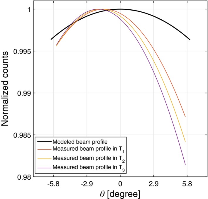
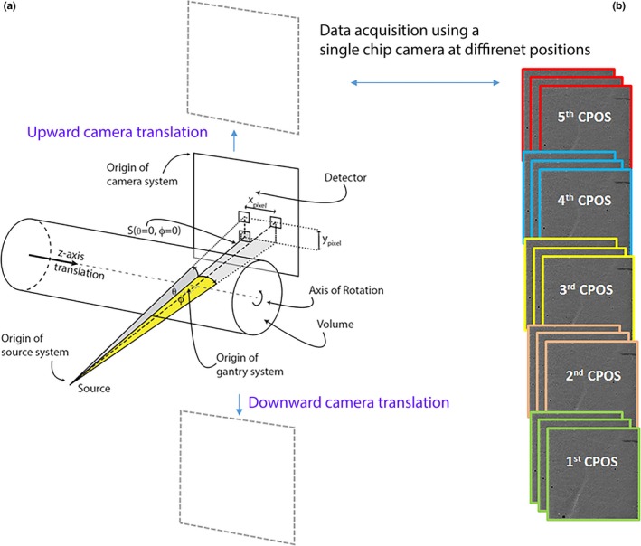
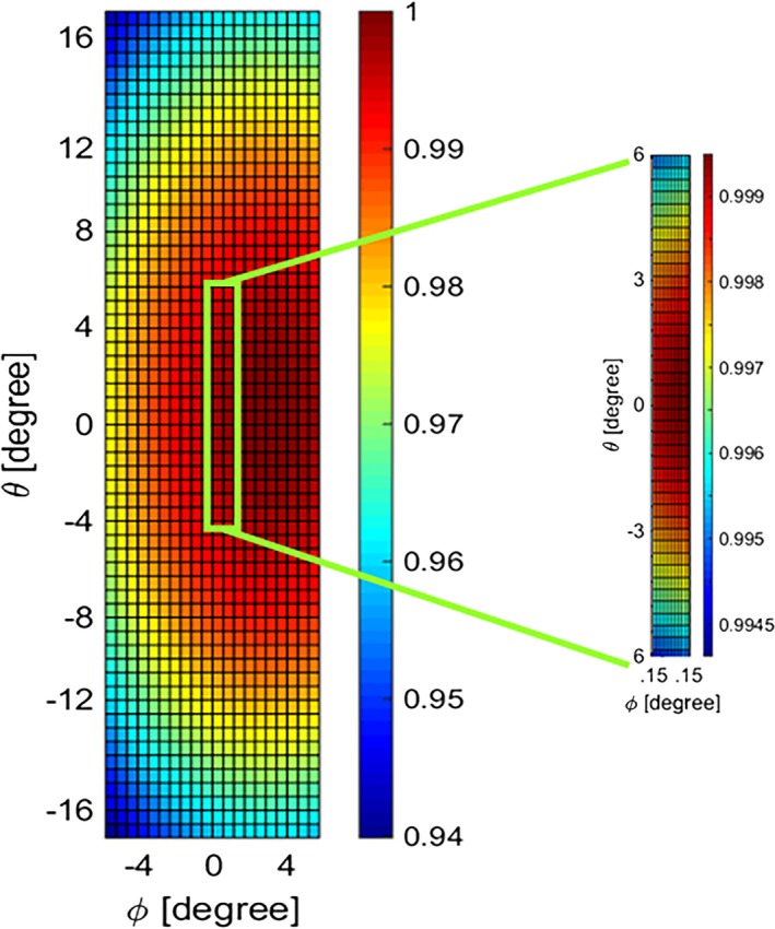
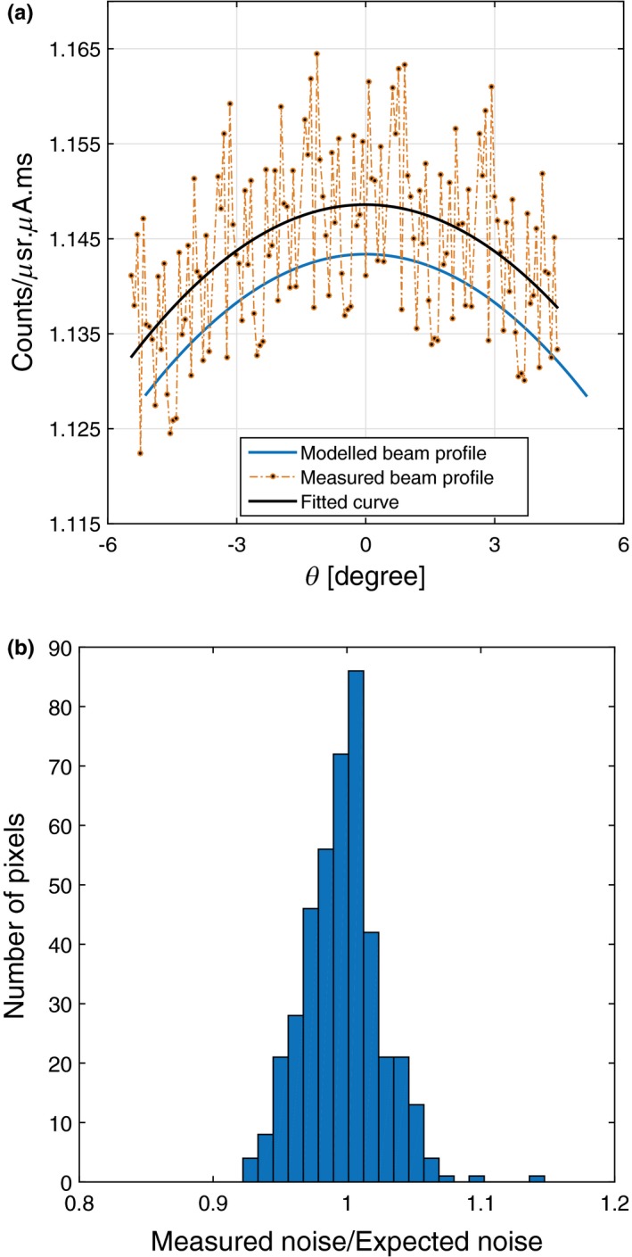
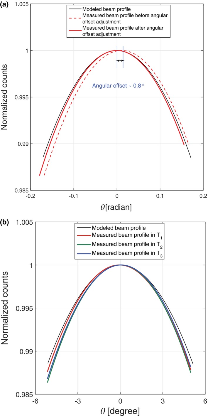
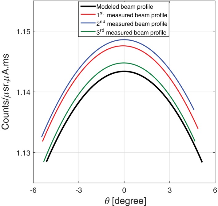
In this paper, we present a method that uses a combination of experimental and modeled data to assess properties of x-ray beam measured using a small-animal spectral scanner. The spatial properties of the beam profile are characterized by beam profile shape, the angular offset along the rotational axis, and the photon count difference between experimental and modeled data at the central beam axis. Temporal stability of the beam profile is assessed by measuring intra- and interscan count variations. The beam profile assessment method was evaluated on several spectral CT scanners equipped with Medipix3RX-based detectors. On a well-calibrated spectral CT scanner, we measured an integral count error of 0.5%, intrascan count variation of 0.1%, and an interscan count variation of less than 1%. The angular offset of the beam center ranged from 0.8° to 1.6° for the studied spectral CT scanners. We also demonstrate the capability of this method to identify poor performance of the system through analyzing the deviation of the experimental beam profile from the model. This technique can, therefore, aid in monitoring the system performance to obtain a robust spectral CT; providing the reliable quantitative images. Furthermore, the accurate offset parameters of a spectral scanner provided by this method allow us to incorporate a more realistic form of the photon distribution in the polychromatic-based image reconstruction models. Both improvements of the reliability of the system and accuracy of the volume reconstruction result in a better discrimination and quantification of the imaged materials.

摘要

在本文中,我们提出了一种使用实验和建模数据相结合的方法,用于评估使用小型动物能谱扫描仪测量的 X 射线束的特性。光束轮廓的空间特性由光束轮廓形状、沿旋转轴的角度偏移以及在中心光束轴处实验数据和模型数据之间的光子计数差异来描述。通过测量内部和外部扫描计数变化来评估光束轮廓的时间稳定性。我们在配备基于 Medipix3RX 的探测器的几台光谱 CT 扫描仪上评估了光束轮廓评估方法。在经过良好校准的光谱 CT 扫描仪上,我们测量到积分计数误差为 0.5%,扫描内计数变化为 0.1%,扫描间计数变化小于 1%。研究的光谱 CT 扫描仪的光束中心的角度偏移范围为 0.8°至 1.6°。我们还通过分析实验光束轮廓与模型的偏差,展示了该方法识别系统性能不佳的能力。因此,该技术可以帮助监测系统性能,以获得稳健的光谱 CT,提供可靠的定量图像。此外,该方法提供的光谱扫描仪的准确偏移参数允许我们在基于多色的图像重建模型中加入更真实的光子分布形式。系统可靠性和体积重建准确性的这两个改进都可以更好地区分和量化成像材料。

https://cdn.ncbi.nlm.nih.gov/pmc/blobs/ae40/5849855/b1e39f282989/ACM2-19-287-g011.jpg
https://cdn.ncbi.nlm.nih.gov/pmc/blobs/ae40/5849855/cbbc73e5db34/ACM2-19-287-g001.jpg
https://cdn.ncbi.nlm.nih.gov/pmc/blobs/ae40/5849855/a31d6547d6d6/ACM2-19-287-g002.jpg
https://cdn.ncbi.nlm.nih.gov/pmc/blobs/ae40/5849855/5ac96758bf0c/ACM2-19-287-g003.jpg
https://cdn.ncbi.nlm.nih.gov/pmc/blobs/ae40/5849855/22027b29a649/ACM2-19-287-g004.jpg
https://cdn.ncbi.nlm.nih.gov/pmc/blobs/ae40/5849855/8398e73ecef3/ACM2-19-287-g005.jpg
https://cdn.ncbi.nlm.nih.gov/pmc/blobs/ae40/5849855/d1c7bdc2109c/ACM2-19-287-g006.jpg
https://cdn.ncbi.nlm.nih.gov/pmc/blobs/ae40/5849855/32463b6976f4/ACM2-19-287-g007.jpg
https://cdn.ncbi.nlm.nih.gov/pmc/blobs/ae40/5849855/22036b49f297/ACM2-19-287-g008.jpg
https://cdn.ncbi.nlm.nih.gov/pmc/blobs/ae40/5849855/9d02837b99c1/ACM2-19-287-g009.jpg
https://cdn.ncbi.nlm.nih.gov/pmc/blobs/ae40/5849855/97a2eb30d076/ACM2-19-287-g010.jpg
https://cdn.ncbi.nlm.nih.gov/pmc/blobs/ae40/5849855/b1e39f282989/ACM2-19-287-g011.jpg

相似文献

1
Beam profile assessment in spectral CT scanners.光谱 CT 扫描仪中的射束轮廓评估。
J Appl Clin Med Phys. 2018 Mar;19(2):287-297. doi: 10.1002/acm2.12260. Epub 2018 Feb 7.
2
Accurate technique for complete geometric calibration of cone-beam computed tomography systems.用于锥束计算机断层扫描系统完全几何校准的精确技术。
Med Phys. 2005 Apr;32(4):968-83. doi: 10.1118/1.1869652.
3
Empirical binary tomography calibration (EBTC) for the precorrection of beam hardening and scatter for flat panel CT.用于平板 CT 束硬化和散射预校正的经验二进制断层摄影术校准 (EBTC)。
Med Phys. 2011 Apr;38(4):2233-40. doi: 10.1118/1.3561506.
4
Assessment of longitudinal beam property and contrast uniformity for 256- and 320-row area detector computed tomography scanners in the 160-mm nonhelical volume-acquisition mode.评估 160mm 非螺旋容积采集模式下 256 排和 320 排探测器 CT 扫描仪的纵向射束性能和对比度均匀性。
J Appl Clin Med Phys. 2019 Aug;20(8):164-170. doi: 10.1002/acm2.12670. Epub 2019 Jun 29.
5
Effects of ray profile modeling on resolution recovery in clinical CT.射线轮廓建模对临床 CT 中分辨率恢复的影响。
Med Phys. 2014 Feb;41(2):021907. doi: 10.1118/1.4862510.
6
Chord-based image reconstruction in cone-beam CT with a curved detector.基于弦的弯曲探测器锥束CT图像重建
Med Phys. 2006 Oct;33(10):3743-57. doi: 10.1118/1.2337270.
7
Multi-slice helical CT: scan and reconstruction.多层螺旋CT:扫描与重建。
Med Phys. 1999 Jan;26(1):5-18. doi: 10.1118/1.598470.
8
Comparison of dosimetric properties among four commercial multi-detector computed tomography scanners.四种商用多探测器计算机断层扫描扫描仪的剂量学特性比较。
Phys Med. 2017 Mar;35:50-58. doi: 10.1016/j.ejmp.2017.02.007. Epub 2017 Feb 21.
9
Quantitative image-based spectral reconstruction for computed tomography.基于定量图像的计算机断层扫描光谱重建。
Med Phys. 2009 Oct;36(10):4471-85. doi: 10.1118/1.3213534.
10
Flat panel detector-based cone beam computed tomography with a circle-plus-two-arcs data acquisition orbit: preliminary phantom study.基于平板探测器的采用圆加双弧数据采集轨道的锥束计算机断层扫描:初步体模研究
Med Phys. 2003 Jul;30(7):1694-705. doi: 10.1118/1.1582470.

引用本文的文献

1
Gold Nanoparticle Mediated Multi-Modal CT Imaging of Hsp70 Membrane-Positive Tumors.金纳米颗粒介导的热休克蛋白70膜阳性肿瘤的多模态CT成像
Cancers (Basel). 2020 May 22;12(5):1331. doi: 10.3390/cancers12051331.
2
Evaluation of a photon counting Medipix3RX cadmium zinc telluride spectral x-ray detector.光子计数型Medipix3RX碲锌镉光谱X射线探测器的评估
J Med Imaging (Bellingham). 2018 Oct;5(4):043503. doi: 10.1117/1.JMI.5.4.043503. Epub 2018 Dec 4.

本文引用的文献

1
Evaluation of a photon counting Medipix3RX CZT spectral x-ray detector.光子计数Medipix3RX CZT光谱X射线探测器的评估。
Proc SPIE Int Soc Opt Eng. 2016 Aug 28;9969. doi: 10.1117/12.2236501. Epub 2016 Oct 3.
2
Quantitative imaging of excised osteoarthritic cartilage using spectral CT.使用光谱CT对切除的骨关节炎软骨进行定量成像。
Eur Radiol. 2017 Jan;27(1):384-392. doi: 10.1007/s00330-016-4374-7. Epub 2016 May 10.
3
Allowable forward model misspecification for accurate basis decomposition in a silicon detector based spectral CT.
硅基探测器能谱 CT 中准确基分解的允许正向模型失配。
IEEE Trans Med Imaging. 2015 Mar;34(3):788-95. doi: 10.1109/TMI.2014.2361680. Epub 2014 Oct 9.
4
A cascaded model of spectral distortions due to spectral response effects and pulse pileup effects in a photon-counting x-ray detector for CT.用于CT的光子计数X射线探测器中由于光谱响应效应和脉冲堆积效应引起的光谱失真的级联模型。
Med Phys. 2014 Apr;41(4):041905. doi: 10.1118/1.4866890.
5
Vision 20/20: Single photon counting x-ray detectors in medical imaging.视野 20/20:医学成像中的单光子计数 X 射线探测器。
Med Phys. 2013 Oct;40(10):100901. doi: 10.1118/1.4820371.
6
Hybrid spectral micro-CT: system design, implementation, and preliminary results.混合光谱微 CT:系统设计、实现与初步结果。
IEEE Trans Biomed Eng. 2014 Feb;61(2):246-53. doi: 10.1109/TBME.2013.2279673.
7
Spectral CT of carotid atherosclerotic plaque: comparison with histology.颈动脉粥样硬化斑块的能谱 CT 研究:与组织病理学对照
Eur Radiol. 2012 Dec;22(12):2581-8. doi: 10.1007/s00330-012-2538-7. Epub 2012 Jul 4.
8
Spectroscopic (multi-energy) CT distinguishes iodine and barium contrast material in MICE.光谱(多能量) CT 可区分 MICE 中的碘和钡造影剂。
Eur Radiol. 2010 Sep;20(9):2126-34. doi: 10.1007/s00330-010-1768-9. Epub 2010 Mar 23.
9
Algorithm for X-ray scatter, beam-hardening, and beam profile correction in diagnostic (kilovoltage) and treatment (megavoltage) cone beam CT.诊断(千伏)和治疗(兆伏)锥形束CT中X射线散射、束硬化和束轮廓校正算法
IEEE Trans Med Imaging. 2008 Dec;27(12):1791-810. doi: 10.1109/TMI.2008.928922.