Department of Radiology, Athinoula A. Martinos Center for Biomedical Imaging, Massachusetts General Hospital Harvard Medical School, Boston, MA
Department of Computer Science, Institute of Mathematics and Statistics, University of São Paulo, Brazil.
J Am Heart Assoc. 2018 Feb 2;7(3):e007834. doi: 10.1161/JAHA.117.007834.
Late gadolinium enhancement (LGE) is the current standard for myocardial scar delineation. In this study, we introduce the tractographic propagation angle (PA), a metric of myofiber curvature (degrees/unit distance) derived from diffusion tensor imaging (DTI), and compare its use to LGE and invasive scar assessment by endocardial voltage mapping.
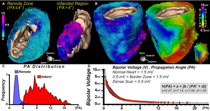
DTI was performed on 7 healthy human volunteers, 5 patients with myocardial infarction, 6 normal mice, and 7 mice with myocardial infarction. LGE to delineate the infarct and border zones was performed with a 2-dimensional inversion recovery gradient-echo sequence. Ex vivo DTI was performed on 5 normal human and 5 normal sheep hearts. Endocardial electroanatomic mapping and subsequent ex vivo DTI was performed on 5 infarcted sheep hearts. PA in the normal human hearts varied smoothly and was generally <4. The mean PA in the infarct zone was significantly elevated (10.34±1.02 versus 4.05±0.45, <0.05). Regions with a PA ≤4 consistently had a bipolar voltage ≥1.5 mV, whereas those with PA values between 4 and 10 had voltages between 0.5 and 1.5 mV. A PA threshold >4 was the most accurate DTI-derived measure of infarct size and demonstrated the greatest correlation with LGE (=0.95).
We found a strong correlation between infarct size by PA and LGE in both mice and humans. There was also an inverse relationship between PA values and endocardial voltage. The use of PA may enable myocardial scar delineation and characterization of arrhythmogenic substrate without the need for exogenous contrast agents.
晚期钆增强(LGE)是目前心肌瘢痕描绘的标准。在这项研究中,我们引入了轨迹传播角(PA),这是一种从弥散张量成像(DTI)中提取的肌纤维曲率度量(度/单位距离),并将其与 LGE 以及心内膜电压测绘的侵入性瘢痕评估进行了比较。
对 7 名健康志愿者、5 名心肌梗死患者、6 只正常小鼠和 7 只心肌梗死小鼠进行了 DTI 检查。使用二维反转恢复梯度回波序列进行 LGE 以描绘梗死区和边界区。在 5 个正常人心和 5 个正常羊心中进行了离体 DTI 检查。在 5 只梗死羊心中进行了心内膜电解剖测绘和随后的离体 DTI 检查。正常人心的 PA 变化平稳,通常<4。梗死区的平均 PA 显著升高(10.34±1.02 比 4.05±0.45,<0.05)。PA 值≤4 的区域始终具有双极电压≥1.5 mV,而 PA 值在 4 到 10 之间的区域的电压在 0.5 到 1.5 mV 之间。PA 值>4 是 DTI 衍生的梗死面积最准确的测量指标,与 LGE 具有最强的相关性(=0.95)。
我们发现小鼠和人类的 PA 与 LGE 之间的梗死面积有很强的相关性。PA 值与心内膜电压之间也存在反比关系。PA 的使用可以在不需要外源性对比剂的情况下实现心肌瘢痕描绘和心律失常基质的特征描述。