• 文献检索
  • 文档翻译
  • 深度研究
  • 学术资讯
  • Suppr Zotero 插件Zotero 插件
  • 邀请有礼
  • 套餐&价格
  • 历史记录
应用&插件
Suppr Zotero 插件Zotero 插件浏览器插件Mac 客户端Windows 客户端微信小程序
定价
高级版会员购买积分包购买API积分包
服务
文献检索文档翻译深度研究API 文档MCP 服务
关于我们
关于 Suppr公司介绍联系我们用户协议隐私条款
关注我们

Suppr 超能文献

核心技术专利:CN118964589B侵权必究
粤ICP备2023148730 号-1Suppr @ 2026

文献检索

告别复杂PubMed语法,用中文像聊天一样搜索,搜遍4000万医学文献。AI智能推荐,让科研检索更轻松。

立即免费搜索

文件翻译

保留排版,准确专业,支持PDF/Word/PPT等文件格式,支持 12+语言互译。

免费翻译文档

深度研究

AI帮你快速写综述,25分钟生成高质量综述,智能提取关键信息,辅助科研写作。

立即免费体验

2型脊髓小脑共济失调中橄榄下核的MRI信号异常

MRI Signal Abnormalities of the Inferior Olivary Nuclei in Spinocerebellar Ataxia Type 2.

作者信息

Yoshii Fumihito, Tomiyasu Hitoshi, Watanabe Ryo, Ryo Masafuchi

机构信息

Department of Neurology, Saiseikai Shonan Hiratsuka Hospital, Hiratsuka, Japan.

Department of Neurology, Tokai University Oiso Hospital, Oiso, Japan.

出版信息

Case Rep Neurol. 2017 Nov 10;9(3):267-271. doi: 10.1159/000481303. eCollection 2017 Sep-Dec.

DOI:10.1159/000481303
PMID:29422848
原文链接:https://pmc.ncbi.nlm.nih.gov/articles/PMC5803720/
Abstract

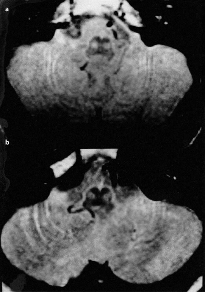
Spinocerebellar ataxia type 2 (SCA2) is an autosomal dominant spinocerebellar degeneration, associated with extended repeats of the trinucleotide CAG in the gene on the long arm of chromosome 12. Magnetic resonance imaging (MRI) of SCA2 showed significant atrophies of the brainstem, middle cerebellar peduncles, and cerebellum. We report two genetically proven SCA2 patients who showed hypertrophy of the inferior olivary nuclei on proton density- and T2-weighted MRI. This pattern has never been reported in patients with SCA1, SCA3, or SCA6, and may make it possible to differentiate SCA2 from other hereditary spinocerebellar ataxias.

摘要

2型脊髓小脑共济失调(SCA2)是一种常染色体显性脊髓小脑变性疾病,与12号染色体长臂上该基因中三核苷酸CAG的重复序列延长有关。SCA2的磁共振成像(MRI)显示脑干、小脑中脚和小脑有明显萎缩。我们报告了两名经基因证实的SCA2患者,他们在质子密度加权和T2加权MRI上显示下橄榄核肥大。这种模式在SCA1、SCA3或SCA6患者中从未有过报道,这可能有助于将SCA2与其他遗传性脊髓小脑共济失调区分开来。

https://cdn.ncbi.nlm.nih.gov/pmc/blobs/5868/5803720/1c3e2a4af8df/crn-0009-0267-g01.jpg

相似文献

1
MRI Signal Abnormalities of the Inferior Olivary Nuclei in Spinocerebellar Ataxia Type 2.2型脊髓小脑共济失调中橄榄下核的MRI信号异常
Case Rep Neurol. 2017 Nov 10;9(3):267-271. doi: 10.1159/000481303. eCollection 2017 Sep-Dec.
2
Frequency of SCA1, SCA2, SCA3/MJD, SCA6, SCA7, and DRPLA CAG trinucleotide repeat expansion in patients with hereditary spinocerebellar ataxia from Chinese kindreds.中国家系遗传性脊髓小脑共济失调患者中SCA1、SCA2、SCA3/MJD、SCA6、SCA7和DRPLA CAG三核苷酸重复扩增的频率
Arch Neurol. 2000 Apr;57(4):540-4. doi: 10.1001/archneur.57.4.540.
3
Autosomal dominant cerebellar ataxia type I. MRI-based volumetry of posterior fossa structures and basal ganglia in spinocerebellar ataxia types 1, 2 and 3.常染色体显性遗传性小脑共济失调I型。基于MRI的1型、2型和3型脊髓小脑共济失调患者后颅窝结构和基底节的容积测定。
Brain. 1998 Sep;121 ( Pt 9):1687-93. doi: 10.1093/brain/121.9.1687.
4
[SCA1, SCA2, MJD/SCA3 (CAG)n mutation detection and analysis in patients with hereditary spinocerebellar ataxia from Chinese families].中国家系遗传性脊髓小脑共济失调患者中SCA1、SCA2、MJD/SCA3(CAG)n突变的检测与分析
Zhonghua Yi Xue Za Zhi. 1997 Nov;77(11):819-22.
5
Spinocerebellar ataxias in Spanish patients: genetic analysis of familial and sporadic cases. The Ataxia Study Group.西班牙患者的脊髓小脑共济失调:家族性和散发性病例的基因分析。共济失调研究小组。
Hum Genet. 1999 Jun;104(6):516-22. doi: 10.1007/s004390050997.
6
Biological and clinical characteristics of individuals at risk for spinocerebellar ataxia types 1, 2, 3, and 6 in the longitudinal RISCA study: analysis of baseline data.在 RISCA 纵向研究中,脊髓小脑共济失调 1、2、3 和 6 型风险个体的生物学和临床特征:基线数据分析。
Lancet Neurol. 2013 Jul;12(7):650-8. doi: 10.1016/S1474-4422(13)70104-2. Epub 2013 May 22.
7
Conversion of individuals at risk for spinocerebellar ataxia types 1, 2, 3, and 6 to manifest ataxia (RISCA): a longitudinal cohort study.将脊髓小脑共济失调 1、2、3、6 型的风险个体转化为显性共济失调(RISCA):一项纵向队列研究。
Lancet Neurol. 2020 Sep;19(9):738-747. doi: 10.1016/S1474-4422(20)30235-0.
8
Structural and functional MRI abnormalities of cerebellar cortex and nuclei in SCA3, SCA6 and Friedreich's ataxia.脊髓小脑共济失调3型、脊髓小脑共济失调6型及弗里德赖希共济失调中小脑皮质和核团的结构与功能磁共振成像异常
Brain. 2015 May;138(Pt 5):1182-97. doi: 10.1093/brain/awv064. Epub 2015 Mar 28.
9
Differences between spinocerebellar ataxias and multiple system atrophy-cerebellar type on proton magnetic resonance spectroscopy.脊髓小脑共济失调与多系统萎缩-小脑型质子磁共振波谱的差异。
PLoS One. 2012;7(10):e47925. doi: 10.1371/journal.pone.0047925. Epub 2012 Oct 31.
10
Molecular genetics of hereditary spinocerebellar ataxia: mutation analysis of spinocerebellar ataxia genes and CAG/CTG repeat expansion detection in 225 Italian families.遗传性脊髓小脑共济失调的分子遗传学:225个意大利家庭中脊髓小脑共济失调基因的突变分析及CAG/CTG重复序列扩增检测
Arch Neurol. 2004 May;61(5):727-33. doi: 10.1001/archneur.61.5.727.

引用本文的文献

1
The Inferior Cerebellar Peduncle Sign: A Novel Imaging Marker for Differentiating Multiple System Atrophy Cerebellar Type from Spinocerebellar Ataxia.小脑下脚征:一种用于鉴别多系统萎缩小脑型与脊髓小脑共济失调的新型影像学标志物。
AJNR Am J Neuroradiol. 2025 Jun 3;46(6):1223-1230. doi: 10.3174/ajnr.A8623.
2
Increased intrinsic membrane excitability is associated with olivary hypertrophy in spinocerebellar ataxia type 1.内在膜兴奋性增加与1型脊髓小脑共济失调中的橄榄核肥大有关。
Hum Mol Genet. 2024 Dec 6;33(24):2159-2176. doi: 10.1093/hmg/ddae146.
3
Increased intrinsic membrane excitability is associated with hypertrophic olivary degeneration in spinocerebellar ataxia type 1.

本文引用的文献

1
MR Imaging in Spinocerebellar Ataxias: A Systematic Review.脊髓小脑共济失调的磁共振成像:一项系统综述。
AJNR Am J Neuroradiol. 2016 Aug;37(8):1405-12. doi: 10.3174/ajnr.A4760. Epub 2016 May 12.
2
Brain pathology of spinocerebellar ataxias.脊髓小脑共济失调的脑病理学。
Acta Neuropathol. 2012 Jul;124(1):1-21. doi: 10.1007/s00401-012-1000-x. Epub 2012 Jun 9.
3
Spinocerebellar ataxia type 2.2型脊髓小脑共济失调
内在膜兴奋性增加与1型脊髓小脑共济失调中的肥大性橄榄核变性相关。
bioRxiv. 2023 Oct 23:2023.10.23.563657. doi: 10.1101/2023.10.23.563657.
4
Molecular Mechanisms and Therapeutics for Spinocerebellar Ataxia Type 2.脊髓小脑性共济失调 2 型的分子机制与治疗。
Neurotherapeutics. 2019 Oct;16(4):1050-1073. doi: 10.1007/s13311-019-00777-6.
5
Diffusion Tensor Imaging in a Case of Pontine Bleeding Showing Hypertrophic Olivary Degeneration and Cerebellar Ataxia.桥脑出血伴肥大性橄榄核变性和小脑共济失调的弥散张量成像
Case Rep Neurol. 2018 Oct 17;10(3):297-301. doi: 10.1159/000493911. eCollection 2018 Sep-Dec.
Handb Clin Neurol. 2012;103:423-36. doi: 10.1016/B978-0-444-51892-7.00026-7.
4
Quantitative assessment of brain stem and cerebellar atrophy in spinocerebellar ataxia types 3 and 6: impact on clinical status.定量评估脊髓小脑共济失调 3 型和 6 型的脑干和小脑萎缩:对临床状况的影响。
AJNR Am J Neuroradiol. 2011 May;32(5):890-7. doi: 10.3174/ajnr.A2387. Epub 2011 Mar 3.
5
Deranged calcium signaling and neurodegeneration in spinocerebellar ataxia type 2.2型脊髓小脑共济失调中的钙信号紊乱与神经退行性变
J Neurosci. 2009 Jul 22;29(29):9148-62. doi: 10.1523/JNEUROSCI.0660-09.2009.
6
Inferior olivary hypertrophy is uncommon in progressive supranuclear palsy.下橄榄核肥大在进行性核上性麻痹中并不常见。
Acta Neuropathol. 2004 Aug;108(2):143-6. doi: 10.1007/s00401-004-0878-3. Epub 2004 May 15.
7
Autosomal dominant cerebellar ataxia type I. MRI-based volumetry of posterior fossa structures and basal ganglia in spinocerebellar ataxia types 1, 2 and 3.常染色体显性遗传性小脑共济失调I型。基于MRI的1型、2型和3型脊髓小脑共济失调患者后颅窝结构和基底节的容积测定。
Brain. 1998 Sep;121 ( Pt 9):1687-93. doi: 10.1093/brain/121.9.1687.
8
Hypertrophy of the inferior olivary nucleus in patients with progressive supranuclear palsy.进行性核上性麻痹患者下橄榄核肥大
Eur Neurol. 1998;39(2):97-102. doi: 10.1159/000007915.
9
MRI in sporadic olivopontocerebellar atrophy and striatonigral degeneration.散发性橄榄体脑桥小脑萎缩和纹状体黑质变性的磁共振成像
Neurology. 1997 Mar;48(3):790-2. doi: 10.1212/wnl.48.3.790.
10
Cloning of the gene for spinocerebellar ataxia 2 reveals a locus with high sensitivity to expanded CAG/glutamine repeats.脊髓小脑共济失调2型基因的克隆揭示了一个对CAG/谷氨酰胺重复序列扩增高度敏感的基因座。
Nat Genet. 1996 Nov;14(3):285-91. doi: 10.1038/ng1196-285.