Li Xin-Wei, Xu Qiu-Shi, Zhang Ren-He, Yang Wei, Li Yu, Zhang Yu-Ming, Tian Yu, Zhang Min, Wang Zhe, Liu Guo-Wen, Xia Cheng, Li Xiao-Bing
Key Laboratory of Zoonosis, Ministry of Education, College of Veterinary Medicine, Jilin University, 5333 Xi'an Road, Changchun, Jilin, 130062, China.
College of animal science and veterinary medicine, Heilongjiang Bayi Agricultural University, Daqing, 163319, Heilongjiang, China.
BMC Vet Res. 2018 Feb 12;14(1):44. doi: 10.1186/s12917-018-1358-7.
The natural incidence of left displacement of abomasum (LDA) in dairy cows was high. The diagnosis of LDA usually relies on characteristic physical exam findings but that transabdominal ultrasound is a useful technique that has been applied to the diagnosis of gastrointestinal diseases of dairy cows in equivocal cases.
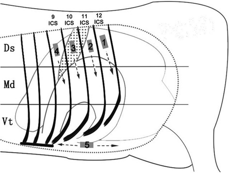
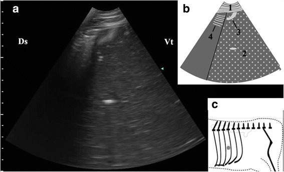
Forty dairy cows with LDA were clinically and ultrasonographically examined to determine the position and the echogenic property of the abomasum. The cows were examined ultrasonographically on the left side, from the 9th intercostal space (ICS) to the 12th ICS as well as the ventral left abdomen before and after reposition surgery.
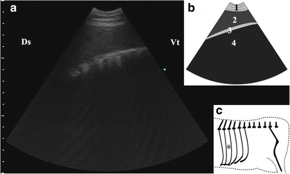
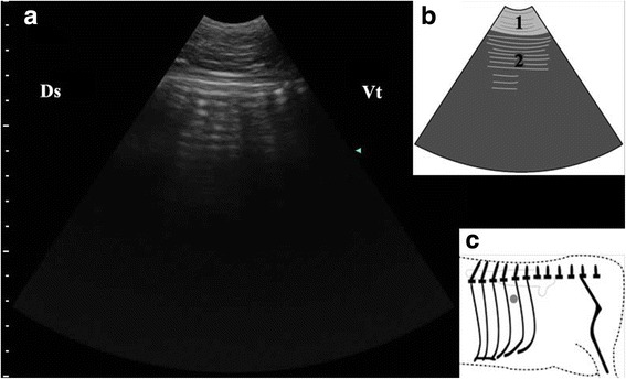
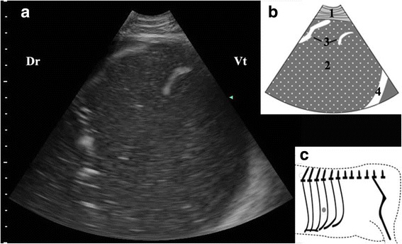
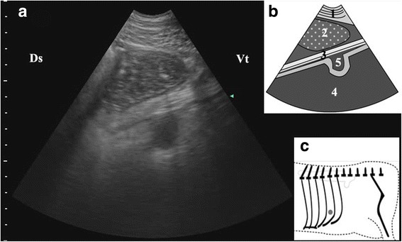
The vital signs were within normal range in most of the cows and the 'pinging' were clearly heard in 37 cows. The abomasal gas cap was visualized from the 9th to 12th ICS in 37 cows and characterized by reverberation artifacts. The abomasal ingesta appeared as homogeneous hypoechoic fluid with scattered hyperechoic foci and were mainly visible in the median region and ventral region of the 9th to 11th ICS in 35 cows. The pyloric canal was detected from the ventral left abdomen wall in 30 cows and appeared as a loop with hypoechogenic wall and echogenic luminal contents in cross section.
These typical ultrasonograms, including reverberation artifacts, homogenous hypoechoic structures, are important diagnostic feature in ultrasonography of LDA. Furthermore, the circular acoustic image structure of the pyloric canal is an important characteristic of LDA, so it can be used as an important diagnostic basis of LDA.
奶牛真胃左方变位(LDA)的自然发生率很高。LDA的诊断通常依赖于特征性的体格检查结果,但经腹超声是一种有用的技术,已应用于奶牛胃肠道疾病的疑难病例诊断。
对40头患有LDA的奶牛进行临床和超声检查,以确定真胃的位置和回声特性。在复位手术前后,对奶牛左侧从第9肋间(ICS)到第12肋间以及左腹腹侧进行超声检查。
大多数奶牛的生命体征在正常范围内,37头奶牛能清晰听到“叩击音”。37头奶牛在第9至12肋间可见真胃气顶,表现为混响伪像。35头奶牛的真胃内容物表现为均匀的低回声液性暗区,内有散在的高回声灶,主要见于第9至11肋间的中部和腹侧区域。30头奶牛从左腹腹侧腹壁检测到幽门管,横断面呈低回声壁和高回声管腔内容物的环状结构。
这些典型的超声图像,包括混响伪像、均匀的低回声结构,是LDA超声检查的重要诊断特征。此外,幽门管的圆形声学图像结构是LDA的重要特征,可作为LDA的重要诊断依据。