• 文献检索
  • 文档翻译
  • 深度研究
  • 学术资讯
  • Suppr Zotero 插件Zotero 插件
  • 邀请有礼
  • 套餐&价格
  • 历史记录
应用&插件
Suppr Zotero 插件Zotero 插件浏览器插件Mac 客户端Windows 客户端微信小程序
定价
高级版会员购买积分包购买API积分包
服务
文献检索文档翻译深度研究API 文档MCP 服务
关于我们
关于 Suppr公司介绍联系我们用户协议隐私条款
关注我们

Suppr 超能文献

核心技术专利:CN118964589B侵权必究
粤ICP备2023148730 号-1Suppr @ 2026

文献检索

告别复杂PubMed语法,用中文像聊天一样搜索,搜遍4000万医学文献。AI智能推荐,让科研检索更轻松。

立即免费搜索

文件翻译

保留排版,准确专业,支持PDF/Word/PPT等文件格式,支持 12+语言互译。

免费翻译文档

深度研究

AI帮你快速写综述,25分钟生成高质量综述,智能提取关键信息,辅助科研写作。

立即免费体验

植入脱矿牙本质基质与周围新形成骨之间界面的三维超微结构分析。

Three-dimensional ultrastructural analysis of the interface between an implanted demineralised dentin matrix and the surrounding newly formed bone.

机构信息

Dental and Oral Medical Centre, Kurume University School of Medicine, Kurume, Fukuoka, Japan.

Department of Dentistry and Oral Surgery, Jyosuikai Imamura Hospital, Tosu, Saga, Japan.

出版信息

Sci Rep. 2018 Feb 12;8(1):2858. doi: 10.1038/s41598-018-21291-3.

DOI:10.1038/s41598-018-21291-3
PMID:29434259
原文链接:https://pmc.ncbi.nlm.nih.gov/articles/PMC5809602/
Abstract

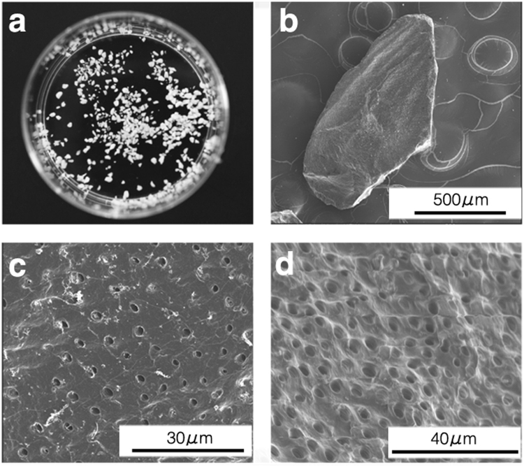
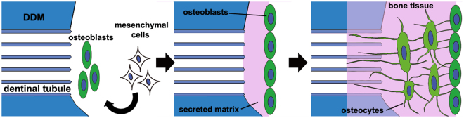
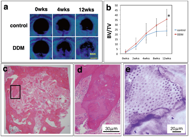
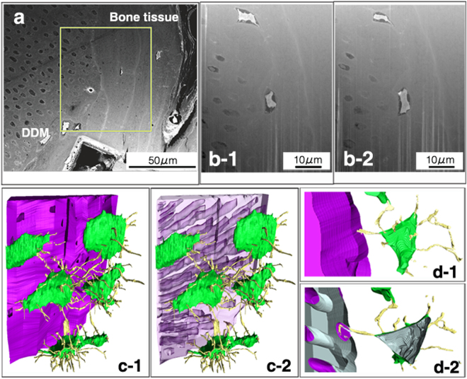
Previous investigators have reported that transplanted demineralised dentin matrix (DDM) influences bone formation in vivo. However, the specific mechanism of how dentinal tubules contribute to bone formation has not been determined with regard to DDM transplantation therapy. In this study, we ultrastructurally investigated how DDM contacted the surrounding newly formed bone using a scanning electron microscopy (SEM) three-dimensional reconstruction method that is based on focused ion beam slicing and SEM (FIB/SEM). A pulverised and processed DDM derived from human teeth was implanted into rat calvarial bone defects, and a series of X-ray computed tomographic images were obtained over 12 weeks. Implants with surrounding new bone were removed and histologically examined using FIB/SEM. After obtaining objective block-face images, the target boundary face was reconstructed three-dimensionally. The osteocytes of the new bone tissue surrounding the DDM formed a network connected by their cellular processes and formed bone tissue. It is also interesting that the cellular processes of the osteocytes extended into the dentinal tubules, and that bone tissue with canaliculi had formed and filled the DDM surface.

摘要

先前的研究人员已经报告说,移植的脱矿牙本质基质(DDM)会影响体内的骨形成。然而,关于 DDM 移植治疗,牙本质小管如何有助于骨形成的具体机制尚不清楚。在这项研究中,我们使用基于聚焦离子束切片和扫描电子显微镜(FIB/SEM)的扫描电子显微镜(SEM)三维重建方法,从超微结构上研究了 DDM 如何与周围新形成的骨接触。将源自人牙齿的粉碎和加工的 DDM 植入大鼠颅骨骨缺损中,并在 12 周内获得一系列 X 射线计算机断层扫描图像。取出带有周围新骨的植入物,并使用 FIB/SEM 进行组织学检查。获得目标块面图像后,对目标边界面进行三维重建。DDM 周围新骨组织中的成骨细胞通过细胞突起形成一个网络,并形成骨组织。有趣的是,成骨细胞的细胞突起延伸到牙本质小管中,并且已经形成并填充了 DDM 表面的有管腔的骨组织。

https://cdn.ncbi.nlm.nih.gov/pmc/blobs/d31d/5809602/f781bf0c2f36/41598_2018_21291_Fig6_HTML.jpg
https://cdn.ncbi.nlm.nih.gov/pmc/blobs/d31d/5809602/21e158352ecb/41598_2018_21291_Fig1_HTML.jpg
https://cdn.ncbi.nlm.nih.gov/pmc/blobs/d31d/5809602/c5f365344780/41598_2018_21291_Fig2_HTML.jpg
https://cdn.ncbi.nlm.nih.gov/pmc/blobs/d31d/5809602/3656655460e1/41598_2018_21291_Fig3_HTML.jpg
https://cdn.ncbi.nlm.nih.gov/pmc/blobs/d31d/5809602/471fa2961664/41598_2018_21291_Fig4_HTML.jpg
https://cdn.ncbi.nlm.nih.gov/pmc/blobs/d31d/5809602/ed3161fcc295/41598_2018_21291_Fig5_HTML.jpg
https://cdn.ncbi.nlm.nih.gov/pmc/blobs/d31d/5809602/f781bf0c2f36/41598_2018_21291_Fig6_HTML.jpg

相似文献

1
Three-dimensional ultrastructural analysis of the interface between an implanted demineralised dentin matrix and the surrounding newly formed bone.植入脱矿牙本质基质与周围新形成骨之间界面的三维超微结构分析。
Sci Rep. 2018 Feb 12;8(1):2858. doi: 10.1038/s41598-018-21291-3.
2
Three-dimensional ultrastructure of osteocytes assessed by focused ion beam-scanning electron microscopy (FIB-SEM).通过聚焦离子束扫描电子显微镜(FIB-SEM)评估骨细胞的三维超微结构。
Histochem Cell Biol. 2018 Apr;149(4):423-432. doi: 10.1007/s00418-018-1645-1. Epub 2018 Feb 9.
3
Evaluation of perforated demineralized dentin scaffold on bone regeneration in critical-size sheep iliac defects.评价脱矿牙本质多孔支架在绵羊临界尺寸髂骨缺损骨再生中的作用。
Clin Oral Implants Res. 2017 Nov;28(11):e227-e235. doi: 10.1111/clr.13000. Epub 2017 Jan 17.
4
Demineralized dentin matrix acts as a scaffold for repair of articular cartilage defects.脱矿牙本质基质可作为关节软骨缺损修复的支架。
Calcif Tissue Int. 2009 Mar;84(3):210-20. doi: 10.1007/s00223-008-9205-7. Epub 2009 Jan 29.
5
Clinical application of autogenous demineralized dentin matrix loaded with recombinant human bone morphogenetic-2 for socket preservation: A case series.自体脱矿牙本质基质负载重组人骨形态发生蛋白-2 用于牙槽窝保存的临床应用:病例系列研究。
Clin Implant Dent Relat Res. 2019 Feb;21(1):4-10. doi: 10.1111/cid.12710. Epub 2018 Dec 27.
6
Demineralized dentin matrix composite collagen material for bone tissue regeneration.脱矿牙本质基质复合胶原材料用于骨组织再生。
J Biomater Sci Polym Ed. 2013;24(13):1519-28. doi: 10.1080/09205063.2013.777227. Epub 2013 Mar 13.
7
Peptide decorated demineralized dentin matrix with enhanced bioactivity, osteogenic differentiation via carboxymethyl chitosan.经羧甲基壳聚糖修饰的具有增强生物活性的脱矿牙本质基质,可通过诱导成骨分化。
Dent Mater. 2021 Jan;37(1):19-29. doi: 10.1016/j.dental.2020.09.019. Epub 2020 Nov 27.
8
Effects of masticatory movement on cranial bone mass and micromorphology of osteocytes and osteoblasts in developing rats.咀嚼运动对发育中大鼠颅骨骨量以及骨细胞和成骨细胞微观形态的影响。
Asia Pac J Clin Nutr. 2009;18(1):96-104.
9
Auto-tooth bone graft material for reconstruction of bone defects in the oral region: case reports.口腔颌骨缺损重建用自体牙骨移植材料:病例报告。
Folia Med (Plovdiv). 2022 Feb 28;64(1):162-168. doi: 10.3897/folmed.64.e59099.
10
The healing capacity and osteogenesis pattern of demineralized dentin matrix (DDM)-fibrin glue (FG) compound.脱矿牙本质基质(DDM)-纤维蛋白胶(FG)复合物的愈合能力和成骨模式。
Sci Rep. 2023 Aug 12;13(1):13140. doi: 10.1038/s41598-023-40258-7.

引用本文的文献

1
A Histologic Study on the Use of Tooth as a Graft Material in Oral Surgery: Analysis of 187 Samples.一项关于牙齿作为口腔外科移植材料应用的组织学研究:187份样本分析
Materials (Basel). 2025 May 27;18(11):2518. doi: 10.3390/ma18112518.
2
Progress in Dentin-Derived Bone Graft Materials: A New Xenogeneic Dentin-Derived Material with Retained Organic Component Allows for Broader and Easier Application.牙本质衍生骨移植材料的研究进展:一种新型保留有机成分的异种牙本质衍生材料,具有更广泛和更容易的应用前景。
Cells. 2024 Oct 31;13(21):1806. doi: 10.3390/cells13211806.
3
Tumor Microenvironment in Oral Squamous Cell Carcinoma: Special Stains and Scanning Electron Microscopic Study.

本文引用的文献

1
Evaluation of perforated demineralized dentin scaffold on bone regeneration in critical-size sheep iliac defects.评价脱矿牙本质多孔支架在绵羊临界尺寸髂骨缺损骨再生中的作用。
Clin Oral Implants Res. 2017 Nov;28(11):e227-e235. doi: 10.1111/clr.13000. Epub 2017 Jan 17.
2
Three-dimensional ultrastructural analyses of anterior pituitary gland expose spatial relationships between endocrine cell secretory granule localization and capillary distribution.三维超微结构分析前垂体腺暴露内分泌细胞分泌颗粒定位和毛细血管分布之间的空间关系。
Sci Rep. 2016 Oct 31;6:36019. doi: 10.1038/srep36019.
3
Histomorphometric and ultrastructural analysis of the tendon-bone interface after rotator cuff repair in a rat model.
口腔鳞状细胞癌中的肿瘤微环境:特殊染色及扫描电子显微镜研究
J Microsc Ultrastruct. 2022 Nov 14;12(3):148-154. doi: 10.4103/jmau.jmau_19_22. eCollection 2024 Jul-Sep.
4
Use of autologous tooth-derived material as a graft in the post-extraction socket. Split-mouth study with radiological and histological analysis.自体牙源性材料在拔牙窝中的应用。放射学和组织学分析的分口研究。
BMC Oral Health. 2024 Jul 23;24(1):832. doi: 10.1186/s12903-024-04576-0.
5
2D vs. 3D Evaluation of Osteocyte Lacunae - Methodological Approaches, Recommended Parameters, and Challenges: A Narrative Review by the European Calcified Tissue Society (ECTS).2D 与 3D 评估破骨细胞陷窝:方法学方法、推荐参数和挑战:欧洲钙化组织学会 (ECTS) 的叙述性综述。
Curr Osteoporos Rep. 2024 Aug;22(4):396-415. doi: 10.1007/s11914-024-00877-z. Epub 2024 Jul 9.
6
A 3D In-vitro model of the human dentine interface shows long-range osteoinduction from the dentine surface.一种 3D 人牙本质界面的体外模型显示,牙本质表面具有远程成骨诱导作用。
Int J Oral Sci. 2024 May 11;16(1):37. doi: 10.1038/s41368-024-00298-9.
7
Demineralized dentin matrix for bone regeneration in dentistry: A critical update.用于牙科骨再生的脱矿牙本质基质:重要更新
Saudi Dent J. 2024 Mar;36(3):443-450. doi: 10.1016/j.sdentj.2023.11.028. Epub 2023 Nov 28.
8
The healing capacity and osteogenesis pattern of demineralized dentin matrix (DDM)-fibrin glue (FG) compound.脱矿牙本质基质(DDM)-纤维蛋白胶(FG)复合物的愈合能力和成骨模式。
Sci Rep. 2023 Aug 12;13(1):13140. doi: 10.1038/s41598-023-40258-7.
9
Autologous Tooth Graft: Innovative Biomaterial for Bone Regeneration. Tooth Transformer and the Role of Microbiota in Regenerative Dentistry. A Systematic Review.自体牙移植:用于骨再生的创新生物材料。牙齿转化器与微生物群在再生牙科中的作用。一项系统评价。
J Funct Biomater. 2023 Feb 27;14(3):132. doi: 10.3390/jfb14030132.
10
Biomineralization-inspired mineralized hydrogel promotes the repair and regeneration of dentin/bone hard tissue.生物矿化启发的矿化水凝胶促进牙本质/骨硬组织的修复和再生。
NPJ Regen Med. 2023 Feb 25;8(1):11. doi: 10.1038/s41536-023-00286-3.
在大鼠模型中修复肩袖后腱骨界面的组织形态计量学和超微结构分析。
Sci Rep. 2016 Sep 20;6:33800. doi: 10.1038/srep33800.
4
Scanning Electron Microscopy and Energy Dispersive X-ray Spectroscopy Studies on Processed Tooth Graft Material by Vacuum-ultrasonic Acceleration.真空超声加速处理的牙齿移植材料的扫描电子显微镜和能量色散X射线光谱研究
Maxillofac Plast Reconstr Surg. 2014 May;36(3):103-10. doi: 10.14402/jkamprs.2014.36.3.103. Epub 2014 May 30.
5
Bone Regeneration Using Dentin Matrix Depends on the Degree of Demineralization and Particle Size.使用牙本质基质进行骨再生取决于脱矿程度和颗粒大小。
PLoS One. 2016 Jan 21;11(1):e0147235. doi: 10.1371/journal.pone.0147235. eCollection 2016.
6
Anchoring structure of the calvarial periosteum revealed by focused ion beam/scanning electron microscope tomography.聚焦离子束/扫描电子显微镜断层扫描揭示的颅盖骨骨膜的锚定结构。
Sci Rep. 2015 Dec 2;5:17511. doi: 10.1038/srep17511.
7
Clinical evaluation of ridge augmentation using autogenous tooth bone graft material: case series study.使用自体牙骨移植材料进行牙槽嵴增高的临床评估:病例系列研究
J Korean Assoc Oral Maxillofac Surg. 2013 Aug;39(4):156-60. doi: 10.5125/jkaoms.2013.39.4.156. Epub 2013 Aug 23.
8
Tooth-derived bone graft material.牙源性骨移植材料。
J Korean Assoc Oral Maxillofac Surg. 2013 Jun;39(3):103-11. doi: 10.5125/jkaoms.2013.39.3.103. Epub 2013 Jun 25.
9
Fiji: an open-source platform for biological-image analysis.斐济:一个用于生物影像分析的开源平台。
Nat Methods. 2012 Jun 28;9(7):676-82. doi: 10.1038/nmeth.2019.
10
Antibacterial activity of dentine and pulp extracellular matrix extracts.牙本质和牙髓细胞外基质提取物的抗菌活性。
Int Endod J. 2012 Aug;45(8):749-55. doi: 10.1111/j.1365-2591.2012.02031.x. Epub 2012 Mar 14.