Meng Yanhong, Zong Ling, Zhang Ziteng, Han Youdong, Wang Yanhui
Department of Ultrasound, The Affiliated Hospital of Jining Medical University, Jining, Shandong 272029, P.R. China.
Department of Thoracic Surgery, The Affiliated Hospital of Jining Medical University, Jining, Shandong 272029, P.R. China.
Exp Ther Med. 2018 Feb;15(2):1493-1499. doi: 10.3892/etm.2017.5544. Epub 2017 Nov 22.
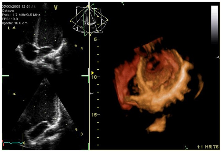
We aimed to evaluate the changes in left ventricular structure and function in hypertensive patients with coronary artery disease before and after percutaneous coronary intervention (PCI) using real-time three-dimensional echocardiography. Two hundred and eighty hypertensive patients with coronary artery disease undergoing PCI and 120 cases who did not receive PCI in our hospital were selected as the subjects of our study. All patients were administered with routine antiplatelet, anticoagulant, lipid-lowering, antihypertensive, dilating coronary artery and other medications. The left ventricular systolic function and systolic synchrony index changes before and after subjects were treated by PCI were analyzed using three-dimensional echocardiography. At 2 days before surgery, there were no significant differences in the left ventricular end-diastolic volume, left ventricular end-systolic volume (LVESV) and ejection fraction (EF) between the two patient groups (P>0.05). At 3 months and 9 months, the two key time points after PCI, the LVESV level in the PCI group was distinctly decreased, while EF was significantly increased (P<0.05). In addition, before treatment, there were no significant differences in the parameters of time from the corresponding segment of the myocardium to the minimal systolic volume in two patient groups, such as Tmsv-16SD, Tmsv-16Dif, Tmsv-12SD, Tmsv-12Dif, Tmsv-6SD and Tmsv-6Dif (P>0.05); however, the parameters of time from the corresponding segment of the myocardium to the minimal systolic volume in patients in the PCI group were significantly reduced at 3 and 9 months after surgery (P<0.05). Three-dimensional echocardiography can evaluate the critical parameters in the prognosis of hypertensive patients with coronary artery disease after PCI accurately and in real-time, which may play a significant role.
我们旨在利用实时三维超声心动图评估冠心病高血压患者经皮冠状动脉介入治疗(PCI)前后左心室结构和功能的变化。选取我院280例接受PCI的冠心病高血压患者和120例未接受PCI的患者作为研究对象。所有患者均给予常规抗血小板、抗凝、降脂、降压、扩张冠状动脉等药物治疗。采用三维超声心动图分析PCI治疗前后受试者左心室收缩功能及收缩同步指数的变化。术前2天,两组患者的左心室舒张末期容积、左心室收缩末期容积(LVESV)和射血分数(EF)比较,差异无统计学意义(P>0.05)。在PCI术后3个月和9个月这两个关键时间点,PCI组的LVESV水平明显降低,而EF显著升高(P<0.05)。此外,治疗前,两组患者心肌相应节段至最小收缩容积时间的参数,如Tmsv-16SD、Tmsv-16Dif、Tmsv-12SD、Tmsv-12Dif、Tmsv-6SD和Tmsv-6Dif比较,差异无统计学意义(P>0.05);但PCI组患者术后3个月和9个月心肌相应节段至最小收缩容积时间的参数显著降低(P<0.05)。三维超声心动图可准确、实时地评估冠心病高血压患者PCI术后预后的关键参数,可能发挥重要作用。