Computational and Systems Biology, John Innes Centre, Norwich NR4 7UH, UK.
Theoretical Biology/Bioinformatics, Dept. of Biology, Utrecht University, Padualaan 8, 3584 CH Utrecht, The Netherlands.
Development. 2018 Mar 20;145(6):dev156778. doi: 10.1242/dev.156778.
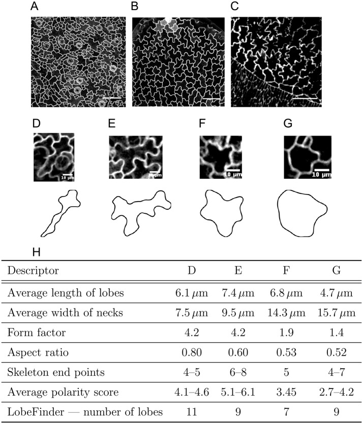
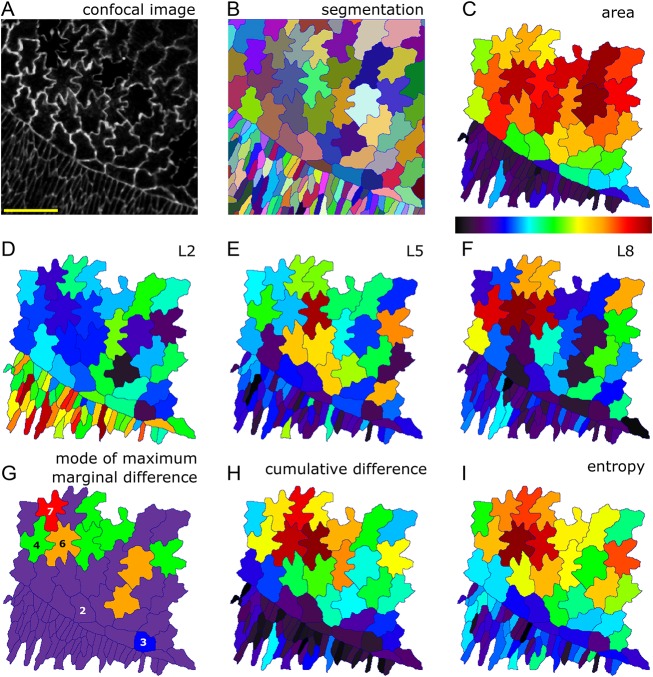
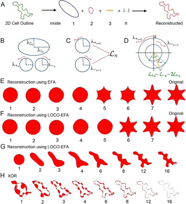
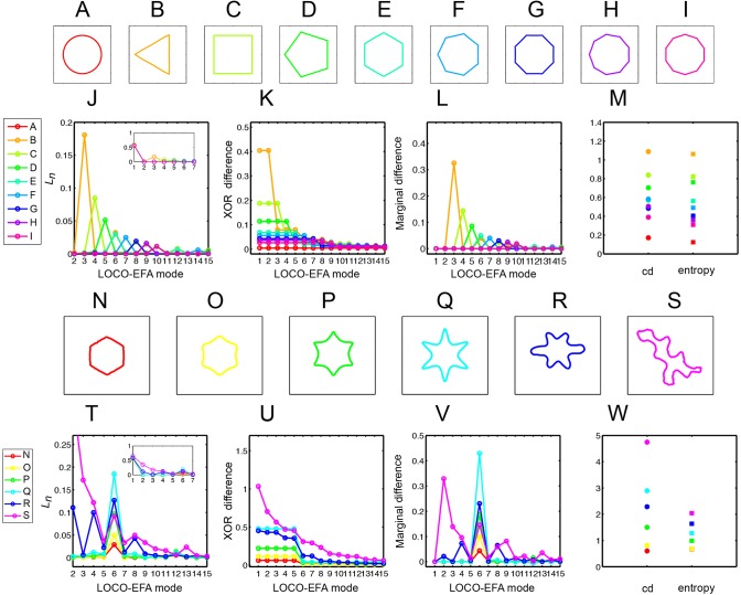
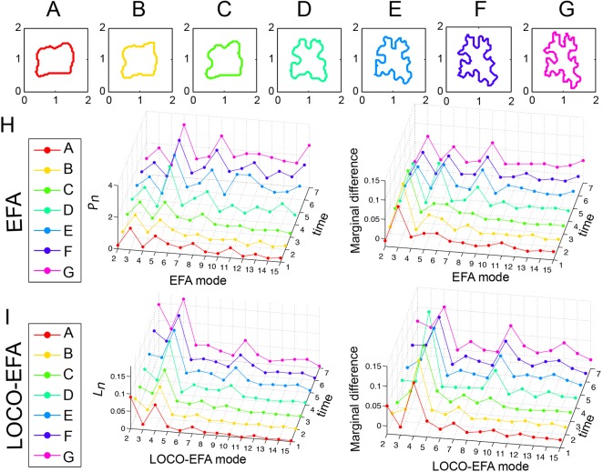
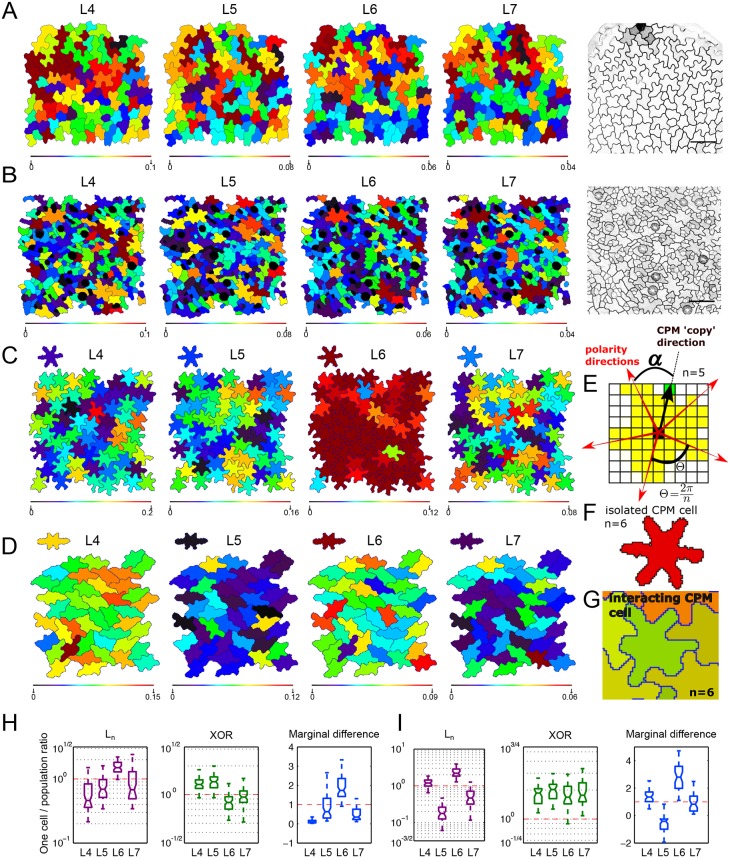
Quantifying cell morphology is fundamental to the statistical study of cell populations, and can help unravel mechanisms underlying cell and tissue morphogenesis. Current methods, however, require extensive human intervention, are highly parameter sensitive, or produce metrics that are difficult to interpret biologically. We therefore developed a method, lobe contribution elliptical Fourier analysis (LOCO-EFA), which generates from digitalised two-dimensional cell outlines meaningful descriptors that can be directly matched to morphological features. This is shown by studying well-defined geometric shapes as well as actual biological cells from plant and animal tissues. LOCO-EFA provides a tool to phenotype efficiently and objectively populations of cells, here demonstrated by applying it to the complex shaped pavement cells of wild-type and leaves, and amnioserosa cells. To validate our method's applicability to large populations, we analysed computer-generated tissues. By controlling cell shape, we explored the potential impact of cell packing on individual cell shape, quantifying through LOCO-EFA deviations between the specified shape of single cells in isolation and the resultant shape when they interact within a confluent tissue.
量化细胞形态是对细胞群体进行统计研究的基础,有助于揭示细胞和组织形态发生的机制。然而,目前的方法需要大量的人工干预,对参数高度敏感,或者产生难以在生物学上解释的指标。因此,我们开发了一种方法,叶瓣贡献椭圆傅里叶分析(LOCO-EFA),它可以从数字化的二维细胞轮廓中生成有意义的描述符,可以直接与形态特征相匹配。这一点通过研究定义明确的几何形状以及来自植物和动物组织的实际生物细胞得到了证明。LOCO-EFA 提供了一种高效、客观地表征细胞群体的工具,在这里我们将其应用于野生型和叶片的复杂形状的 pavement 细胞以及 amnioserosa 细胞中进行了验证。为了验证我们的方法在大群体中的适用性,我们分析了计算机生成的组织。通过控制细胞形状,我们探索了细胞堆积对单个细胞形状的潜在影响,通过 LOCO-EFA 量化了单个细胞在分离状态下的指定形状与它们在连通组织中相互作用时的形状之间的偏差。