Makarchuk Stanislaw, Beyer Nicolas, Gaiddon Christian, Grange Wilfried, Hébraud Pascal
Université de Strasbourg, IPCMS/CNRS, UMR 7504, 23 rue du Loess, Strasbourg, 67034, France.
Université de Strasbourg, Inserm U1113, 3 avenue Molière, Strasbourg, 67200, France.
Sci Rep. 2018 Feb 14;8(1):3038. doi: 10.1038/s41598-018-21206-2.
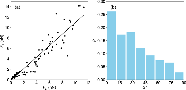
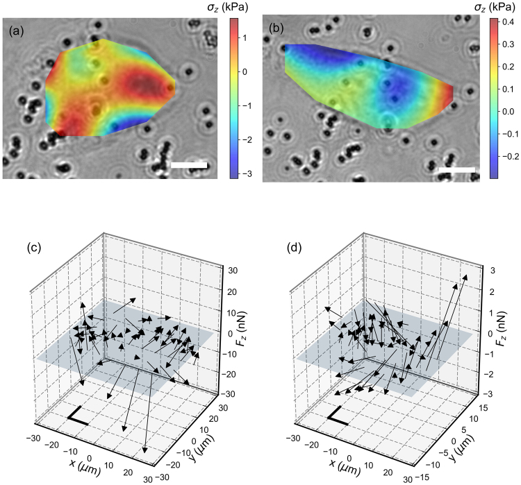
Traction Force Microscopy (TFM) computes the forces exerted at the surface of an elastic material by measuring induced deformations in volume. It is used to determine the pattern of the adhesion forces exerted by cells or by cellular assemblies grown onto a soft deformable substrate. Typically, colloidal particles are dispersed in the substrate and their displacement is monitored by fluorescent microscopy. As with any other fluorescent techniques, the accuracy in measuring a particule's position is ultimately limited by the number of evaluated fluorescent photons. Here, we present a TFM technique based on the detection of probe particle displacements by holographic tracking microscopy. We show that nanometer scale resolutions of the particle displacements can be obtained and determine the maximum volume fraction of markers in the substrate. We demonstrate the feasibility of the technique experimentally and measure the three-dimensional force fields exerted by colorectal cancer cells cultivated onto a polyacrylamide gel substrate.
牵引力显微镜(TFM)通过测量体积中的诱导变形来计算施加在弹性材料表面的力。它用于确定细胞或生长在柔软可变形基质上的细胞组件所施加的粘附力模式。通常,胶体颗粒分散在基质中,其位移通过荧光显微镜进行监测。与任何其他荧光技术一样,测量颗粒位置的准确性最终受评估的荧光光子数量限制。在此,我们提出一种基于全息跟踪显微镜检测探针颗粒位移的TFM技术。我们表明可以获得颗粒位移的纳米级分辨率,并确定基质中标记物的最大体积分数。我们通过实验证明了该技术的可行性,并测量了培养在聚丙烯酰胺凝胶基质上的结肠癌细胞所施加的三维力场。