• 文献检索
  • 文档翻译
  • 深度研究
  • 学术资讯
  • Suppr Zotero 插件Zotero 插件
  • 邀请有礼
  • 套餐&价格
  • 历史记录
应用&插件
Suppr Zotero 插件Zotero 插件浏览器插件Mac 客户端Windows 客户端微信小程序
定价
高级版会员购买积分包购买API积分包
服务
文献检索文档翻译深度研究API 文档MCP 服务
关于我们
关于 Suppr公司介绍联系我们用户协议隐私条款
关注我们

Suppr 超能文献

核心技术专利:CN118964589B侵权必究
粤ICP备2023148730 号-1Suppr @ 2026

文献检索

告别复杂PubMed语法,用中文像聊天一样搜索,搜遍4000万医学文献。AI智能推荐,让科研检索更轻松。

立即免费搜索

文件翻译

保留排版,准确专业,支持PDF/Word/PPT等文件格式,支持 12+语言互译。

免费翻译文档

深度研究

AI帮你快速写综述,25分钟生成高质量综述,智能提取关键信息,辅助科研写作。

立即免费体验

用荧光显微镜和基于对象的空间统计学绘制分子组装图。

Mapping molecular assemblies with fluorescence microscopy and object-based spatial statistics.

机构信息

Institut Pasteur, BioImage Analysis Unit. CNRS UMR 3691. 25 rue du Docteur Roux, 75724, Paris Cedex 15, France.

Department of Biological Sciences, Columbia University, New York, NY, USA.

出版信息

Nat Commun. 2018 Feb 15;9(1):698. doi: 10.1038/s41467-018-03053-x.

DOI:10.1038/s41467-018-03053-x
PMID:29449608
原文链接:https://pmc.ncbi.nlm.nih.gov/articles/PMC5814551/
Abstract

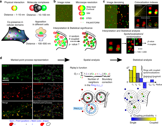
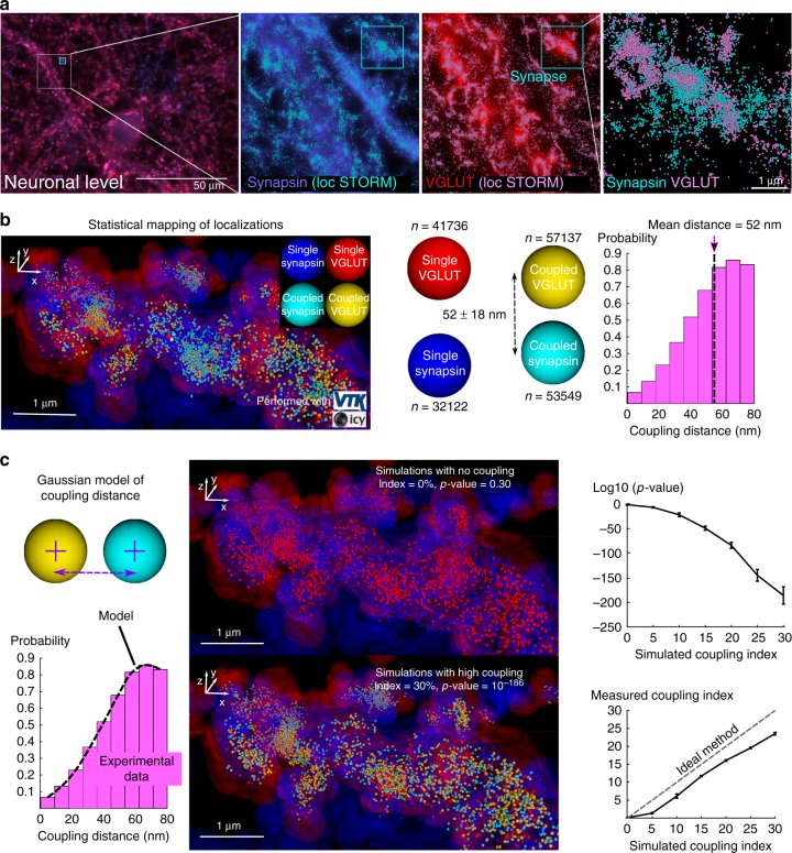
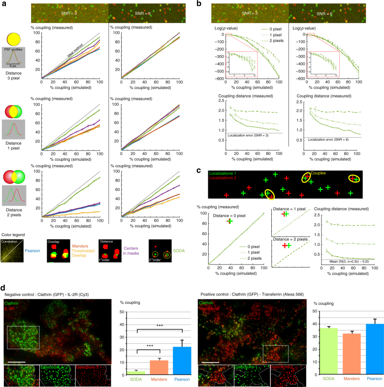
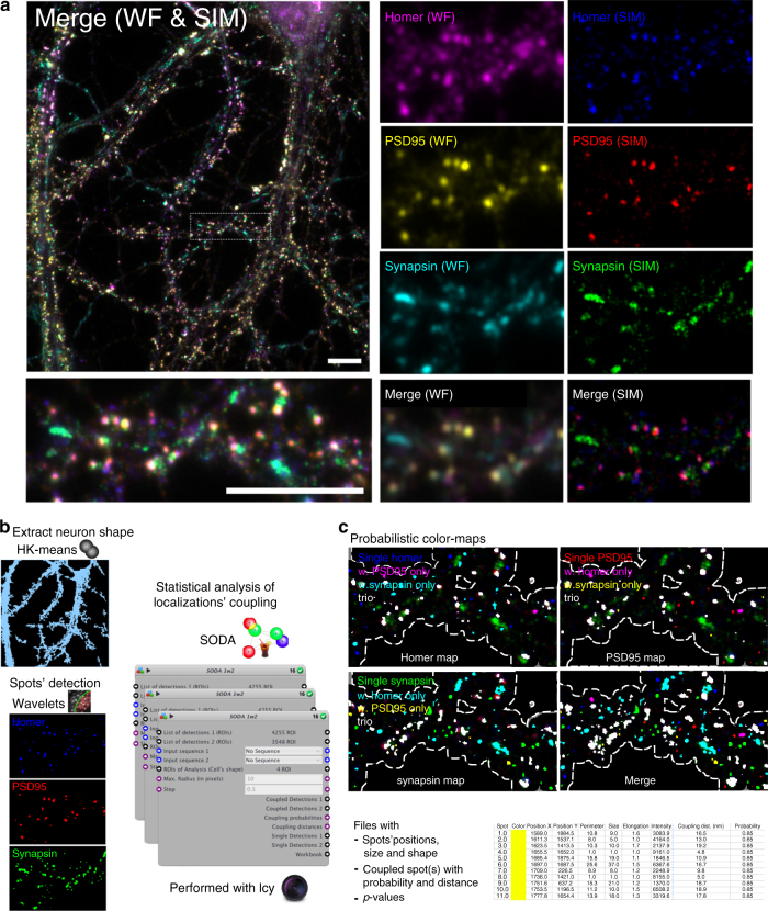
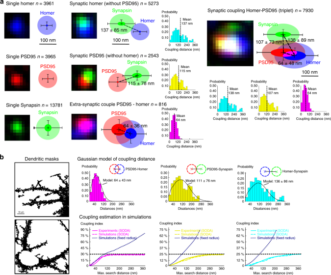
Elucidating protein functions and molecular organisation requires to localise precisely single or aggregated molecules and analyse their spatial distributions. We develop a statistical method SODA (Statistical Object Distance Analysis) that uses either micro- or nanoscopy to significantly improve on standard co-localisation techniques. Our method considers cellular geometry and densities of molecules to provide statistical maps of isolated and associated (coupled) molecules. We use SODA with three-colour structured-illumination microscopy (SIM) images of hippocampal neurons, and statistically characterise spatial organisation of thousands of synapses. We show that presynaptic synapsin is arranged in asymmetric triangle with the 2 postsynaptic markers homer and PSD95, indicating a deeper localisation of homer. We then determine stoichiometry and distance between localisations of two synaptic vesicle proteins with 3D-STORM. These findings give insights into the protein organisation at the synapse, and prove the efficiency of SODA to quantitatively assess the geometry of molecular assemblies.

摘要

阐明蛋白质的功能和分子组织需要精确定位单个或聚集的分子,并分析它们的空间分布。我们开发了一种统计方法 SODA(统计对象距离分析),该方法使用显微镜或纳米显微镜,显著改进了标准共定位技术。我们的方法考虑了细胞的几何形状和分子的密度,为分离和关联(耦合)分子提供了统计图谱。我们使用 SODA 对海马神经元的三色结构光照显微镜 (SIM) 图像进行处理,并从统计学上对数千个突触的空间组织进行了特征描述。我们发现,突触前突触素以不对称三角形的形式排列,与 2 个突触后标记物 homer 和 PSD95 相连,这表明 homer 的定位更深。然后,我们使用 3D-STORM 确定了两个突触小泡蛋白定位之间的化学计量和距离。这些发现为突触处的蛋白质组织提供了深入的了解,并证明了 SODA 定量评估分子组装几何形状的效率。

https://cdn.ncbi.nlm.nih.gov/pmc/blobs/8a0d/5814551/d397c8f64a39/41467_2018_3053_Fig5_HTML.jpg
https://cdn.ncbi.nlm.nih.gov/pmc/blobs/8a0d/5814551/83d491823e8c/41467_2018_3053_Fig1_HTML.jpg
https://cdn.ncbi.nlm.nih.gov/pmc/blobs/8a0d/5814551/720d8babfebd/41467_2018_3053_Fig2_HTML.jpg
https://cdn.ncbi.nlm.nih.gov/pmc/blobs/8a0d/5814551/5d79b416a443/41467_2018_3053_Fig3_HTML.jpg
https://cdn.ncbi.nlm.nih.gov/pmc/blobs/8a0d/5814551/fb53c774d165/41467_2018_3053_Fig4_HTML.jpg
https://cdn.ncbi.nlm.nih.gov/pmc/blobs/8a0d/5814551/d397c8f64a39/41467_2018_3053_Fig5_HTML.jpg

相似文献

1
Mapping molecular assemblies with fluorescence microscopy and object-based spatial statistics.用荧光显微镜和基于对象的空间统计学绘制分子组装图。
Nat Commun. 2018 Feb 15;9(1):698. doi: 10.1038/s41467-018-03053-x.
2
Activity-Dependent Remodeling of Synaptic Protein Organization Revealed by High Throughput Analysis of STED Nanoscopy Images.基于超高分辨 STED 显微镜图像高通量分析揭示的突触蛋白组织的活性依赖性重塑。
Front Neural Circuits. 2020 Oct 15;14:57. doi: 10.3389/fncir.2020.00057. eCollection 2020.
3
Transsynaptic Assemblies Link Domains of Presynaptic and Postsynaptic Intracellular Structures across the Synaptic Cleft.突触前和突触后细胞内结构的域通过突触裂隙的突触传递连接。
J Neurosci. 2023 Aug 16;43(33):5883-5892. doi: 10.1523/JNEUROSCI.2195-22.2023. Epub 2023 Jun 27.
4
Molecular mapping of neuronal architecture using STORM microscopy and new fluorescent probes for SMLM imaging.使用STORM显微镜和用于超分辨光学显微镜成像的新型荧光探针进行神经元结构的分子图谱绘制。
Neurophotonics. 2024 Jan;11(1):014414. doi: 10.1117/1.NPh.11.1.014414. Epub 2024 Mar 8.
5
PSD95 nanoclusters are postsynaptic building blocks in hippocampus circuits.突触后密度蛋白95(PSD95)纳米簇是海马体回路中的突触后构建模块。
Sci Rep. 2016 Apr 25;6:24626. doi: 10.1038/srep24626.
6
Enhanced synaptic protein visualization by multicolor super-resolution expansion microscopy.通过多色超分辨率扩展显微镜增强突触蛋白可视化。
Neurophotonics. 2023 Oct;10(4):044412. doi: 10.1117/1.NPh.10.4.044412. Epub 2023 Oct 25.
7
Hippocampal mossy fibers induce assembly and clustering of PSD95-containing postsynaptic densities independent of glutamate receptor activation.海马苔藓纤维可诱导含PSD95的突触后致密物的组装和聚集,且不依赖于谷氨酸受体激活。
J Comp Neurol. 2001 Nov 19;440(3):284-98. doi: 10.1002/cne.1386.
8
Imaging tripartite synapses using super-resolution microscopy.使用超分辨率显微镜对三分叉突触成像。
Methods. 2020 Mar 1;174:81-90. doi: 10.1016/j.ymeth.2019.05.024. Epub 2019 May 31.
9
The distribution of synapsin I and synaptophysin in hippocampal neurons developing in culture.培养中发育的海马神经元中突触素I和突触囊泡蛋白的分布。
J Neurosci. 1991 Jun;11(6):1617-26. doi: 10.1523/JNEUROSCI.11-06-01617.1991.
10
Homer 1a gates the induction mechanism for endocannabinoid-mediated synaptic plasticity.荷马 1a 门控内源性大麻素介导的突触可塑性的诱导机制。
J Neurosci. 2010 Feb 24;30(8):3072-81. doi: 10.1523/JNEUROSCI.4603-09.2010.

引用本文的文献

1
Identifying intermolecular interactions in single-molecule localization microscopy.在单分子定位显微镜中识别分子间相互作用。
Proc Natl Acad Sci U S A. 2025 May 20;122(20):e2409426122. doi: 10.1073/pnas.2409426122. Epub 2025 May 12.
2
Statistical analysis of spatial patterns in tumor microenvironment images.肿瘤微环境图像中空间模式的统计分析。
Nat Commun. 2025 Mar 31;16(1):3090. doi: 10.1038/s41467-025-57943-y.
3
Unraveling microglial spatial organization in the developing human brain with DeepCellMap, a deep learning approach coupled with spatial statistics.

本文引用的文献

1
Measurement of co-localization of objects in dual-colour confocal images.双色共聚焦图像中物体共定位的测量。
J Microsc. 1993 Mar;169(3):375-382. doi: 10.1111/j.1365-2818.1993.tb03313.x.
2
Decoupling global biases and local interactions between cell biological variables.解耦细胞生物学变量之间的全局偏差和局部相互作用。
Elife. 2017 Mar 13;6:e22323. doi: 10.7554/eLife.22323.
3
A Bayesian cluster analysis method for single-molecule localization microscopy data.基于贝叶斯聚类分析的单分子定位显微镜数据分析方法
利用深度学习方法DeepCellMap结合空间统计学解析发育中人类大脑中的小胶质细胞空间组织。
Nat Commun. 2025 Feb 13;16(1):1577. doi: 10.1038/s41467-025-56560-z.
4
Ubiquitin-mediated recruitment of the ATG9A-ATG2 lipid transfer complex drives clearance of phosphorylated p62 aggregates.泛素介导的ATG9A-ATG2脂质转移复合物募集驱动磷酸化p62聚集体的清除。
Mol Biol Cell. 2025 Feb 1;36(2):ar20. doi: 10.1091/mbc.E24-03-0101. Epub 2024 Dec 24.
5
Phosphorylation of Optineurin by protein kinase D regulates Parkin-dependent mitophagy.蛋白激酶D介导的视神经病相关蛋白磷酸化调控帕金蛋白依赖性线粒体自噬。
iScience. 2024 Nov 13;27(12):111384. doi: 10.1016/j.isci.2024.111384. eCollection 2024 Dec 20.
6
Bilateral regulation of EGFR activity and local PI(4,5)P dynamics in mammalian cells observed with superresolution microscopy.利用超分辨率显微镜观察到哺乳动物细胞中 EGFR 活性和局部 PI(4,5)P 动力学的双向调节。
Elife. 2024 Nov 8;13:e101652. doi: 10.7554/eLife.101652.
7
MultiMatch: geometry-informed colocalization in multi-color super-resolution microscopy.多匹配:多色超分辨率显微镜中的几何信息共定位。
Commun Biol. 2024 Sep 13;7(1):1139. doi: 10.1038/s42003-024-06772-8.
8
ECLiPSE: a versatile classification technique for structural and morphological analysis of 2D and 3D single-molecule localization microscopy data.ECLiPSE:一种用于二维和三维单分子定位显微镜数据的结构和形态分析的通用分类技术。
Nat Methods. 2024 Oct;21(10):1909-1915. doi: 10.1038/s41592-024-02414-3. Epub 2024 Sep 10.
9
Intracellular spatial transcriptomic analysis toolkit (InSTAnT).细胞内空间转录组分析工具包(InSTAnT)。
Nat Commun. 2024 Sep 6;15(1):7794. doi: 10.1038/s41467-024-49457-w.
10
CoPixie, a novel algorithm for single-particle track colocalization, enables efficient quantification of telomerase dynamics at telomeres.CoPixie 是一种用于单颗粒轨迹共定位的新型算法,能够有效地定量端粒酶在端粒处的动力学。
Nucleic Acids Res. 2024 Sep 9;52(16):9417-9430. doi: 10.1093/nar/gkae669.
Nat Protoc. 2016 Dec;11(12):2499-2514. doi: 10.1038/nprot.2016.149. Epub 2016 Nov 17.
4
Mapping Synaptic Input Fields of Neurons with Super-Resolution Imaging.利用超分辨率成像绘制神经元的突触输入场
Cell. 2015 Oct 8;163(2):493-505. doi: 10.1016/j.cell.2015.08.033. Epub 2015 Oct 1.
5
SR-Tesseler: a method to segment and quantify localization-based super-resolution microscopy data.SR-Tesseler:一种用于分割和量化基于定位的超分辨率显微镜数据的方法。
Nat Methods. 2015 Nov;12(11):1065-71. doi: 10.1038/nmeth.3579. Epub 2015 Sep 7.
6
Biochemical Computation for Spine Structural Plasticity.脊柱结构可塑性的生化计算
Neuron. 2015 Jul 1;87(1):63-75. doi: 10.1016/j.neuron.2015.05.043.
7
Statistical analysis of molecule colocalization in bioimaging.生物成像中分子共定位的统计分析。
Cytometry A. 2015 Jun;87(6):568-79. doi: 10.1002/cyto.a.22629. Epub 2015 Jan 20.
8
Composition of isolated synaptic boutons reveals the amounts of vesicle trafficking proteins.分离的突触小泡的组成揭示了囊泡运输蛋白的含量。
Science. 2014 May 30;344(6187):1023-8. doi: 10.1126/science.1252884.
9
Systematic spatial mapping of proteins at exocytic and endocytic structures.对胞吐和胞吞结构处蛋白质进行系统的空间定位
Mol Biol Cell. 2014 Jul 1;25(13):2084-93. doi: 10.1091/mbc.E14-02-0771. Epub 2014 May 7.
10
Molecular structure, function, and dynamics of clathrin-mediated membrane traffic.网格蛋白介导的膜运输的分子结构、功能及动力学
Cold Spring Harb Perspect Biol. 2014 May 1;6(5):a016725. doi: 10.1101/cshperspect.a016725.