• 文献检索
  • 文档翻译
  • 深度研究
  • 学术资讯
  • Suppr Zotero 插件Zotero 插件
  • 邀请有礼
  • 套餐&价格
  • 历史记录
应用&插件
Suppr Zotero 插件Zotero 插件浏览器插件Mac 客户端Windows 客户端微信小程序
定价
高级版会员购买积分包购买API积分包
服务
文献检索文档翻译深度研究API 文档MCP 服务
关于我们
关于 Suppr公司介绍联系我们用户协议隐私条款
关注我们

Suppr 超能文献

核心技术专利:CN118964589B侵权必究
粤ICP备2023148730 号-1Suppr @ 2026

文献检索

告别复杂PubMed语法,用中文像聊天一样搜索,搜遍4000万医学文献。AI智能推荐,让科研检索更轻松。

立即免费搜索

文件翻译

保留排版,准确专业,支持PDF/Word/PPT等文件格式,支持 12+语言互译。

免费翻译文档

深度研究

AI帮你快速写综述,25分钟生成高质量综述,智能提取关键信息,辅助科研写作。

立即免费体验

第一代和第二代药物洗脱支架内再狭窄的光学相干断层扫描特征有所不同。

Optical coherence tomography characteristics of in-stent restenosis are different between first and second generation drug eluting stents.

作者信息

Kilickesmez Kadriye, Dall'Ara Gianni, Rama-Merchan Juan Carlos, Ghione Matteo, Mattesini Alessio, Vinues Carlos Moreno, Konstantinidis Nikolaos, Pighi Michele, Estevez-Loureiro Rodrigo, Zivelonghi Carlo, Lindsay Alistair C, Secco Gioel G, Foin Nicolas, De Silva Ranil, Di Mario Carlo

机构信息

NIHR Biomedical Research Unit, Royal Brompton Hospital & Harefield NHS Foundation Trust, London, UK.

Department of Clinical and Experimental Medicine, University of Eastern Piedmont, Novara, Italy.

出版信息

Int J Cardiol Heart Vessel. 2014 Mar 19;3:68-74. doi: 10.1016/j.ijchv.2014.03.003. eCollection 2014 Jun.

DOI:10.1016/j.ijchv.2014.03.003
PMID:29450174
原文链接:https://pmc.ncbi.nlm.nih.gov/articles/PMC5801271/
Abstract

AIMS

Characterization of neointimal tissue is essential to understand the pathophysiology of in-stent restenosis (ISR) after drug eluting stent (DES) implantation. Using optical coherence tomography (OCT), we compared the morphologic characteristics of ISR between first and second generation DES.

METHODS AND RESULTS

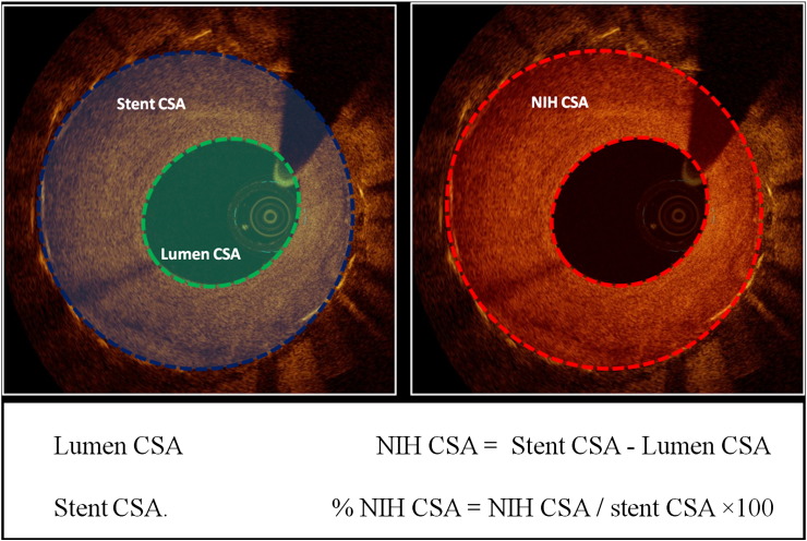
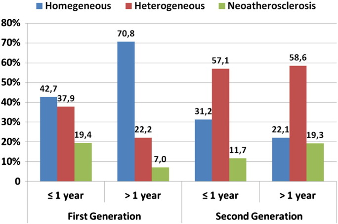
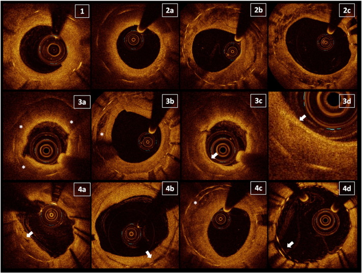
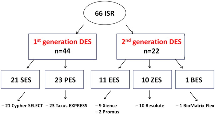
OCT was performed in 66 DES-ISR, defined as > 50% angiographic diameter stenosis within the stented segment. Patients with ISR of first generation sirolimus-eluting stents (SES), paclitaxel eluting stents (PES) and second generation zotarolimus-eluting stents (ZES), everolimus-eluting stents (EES) and biolimus-eluting stents (BES) were enrolled. Quantitative and qualitative ISR tissue analysis was performed at 1-mm intervals along the entire stent, and categorised as homogeneous, heterogeneous and neo-atherosclerosis. The presence of microvessels and peri-strut low intensity area (PSLIA) was determined in all ISR. Neoatherosclerosis was identified by lipid, calcium and thin-cap fibro-atheroma (TCFA) like lesions. We compared the two DES generations at both early (< 1 year) and late (> 1 year) follow-ups.In second generation DES a heterogeneous pattern was prevalent both before and after 1 year (57.1% and 58.6% respectively). Neo-atherosclerosis was more common in the early period in first generation DES (19.4% vs 11.7%, p < 0.01), but after one year was more prevalent in second generation DES (7.0% vs 19.3%, p < 0.01). Similar prevalence of TCFAs was observed in both groups in all comparisons.

CONCLUSIONS

When ISR restenosis occurs in second generation DES, the current data suggest a different time course and different morphological characteristics from first generation. Future prospective studies should evaluate the relationship between ISR morphology, time course and clinical events.

摘要

目的

对新生内膜组织进行特征分析对于理解药物洗脱支架(DES)植入后支架内再狭窄(ISR)的病理生理学至关重要。我们使用光学相干断层扫描(OCT)比较了第一代和第二代DES之间ISR的形态学特征。

方法与结果

对66例DES-ISR患者进行了OCT检查,DES-ISR定义为支架段内血管造影直径狭窄>50%。纳入第一代西罗莫司洗脱支架(SES)、紫杉醇洗脱支架(PES)以及第二代佐他莫司洗脱支架(ZES)、依维莫司洗脱支架(EES)和生物可吸收涂层依维莫司洗脱支架(BES)的ISR患者。沿着整个支架以1毫米的间隔进行ISR组织的定量和定性分析,并分类为均匀型、异质型和新动脉粥样硬化型。在所有ISR中确定微血管和支架周围低强度区域(PSLIA)的存在。通过脂质、钙和薄帽纤维粥样瘤(TCFA)样病变识别新动脉粥样硬化。我们在早期(<1年)和晚期(>1年)随访中比较了两代DES。在第二代DES中,异质型在1年前后均很普遍(分别为57.1%和58.6%)。新动脉粥样硬化在第一代DES的早期更常见(19.4%对11.7%,p<0.01),但1年后在第二代DES中更普遍(7.0%对19.3%,p<0.01)。在所有比较中,两组中TCFAs的发生率相似。

结论

当第二代DES发生ISR再狭窄时,目前的数据表明其时间进程和形态学特征与第一代不同。未来的前瞻性研究应评估ISR形态、时间进程与临床事件之间的关系。

https://cdn.ncbi.nlm.nih.gov/pmc/blobs/2190/5801271/fac0f6ac8dd5/gr4.jpg
https://cdn.ncbi.nlm.nih.gov/pmc/blobs/2190/5801271/7d433c81c2c2/gr1.jpg
https://cdn.ncbi.nlm.nih.gov/pmc/blobs/2190/5801271/b4b75e9e4bd6/gr2.jpg
https://cdn.ncbi.nlm.nih.gov/pmc/blobs/2190/5801271/4db8db38dd68/gr3.jpg
https://cdn.ncbi.nlm.nih.gov/pmc/blobs/2190/5801271/fac0f6ac8dd5/gr4.jpg
https://cdn.ncbi.nlm.nih.gov/pmc/blobs/2190/5801271/7d433c81c2c2/gr1.jpg
https://cdn.ncbi.nlm.nih.gov/pmc/blobs/2190/5801271/b4b75e9e4bd6/gr2.jpg
https://cdn.ncbi.nlm.nih.gov/pmc/blobs/2190/5801271/4db8db38dd68/gr3.jpg
https://cdn.ncbi.nlm.nih.gov/pmc/blobs/2190/5801271/fac0f6ac8dd5/gr4.jpg

相似文献

1
Optical coherence tomography characteristics of in-stent restenosis are different between first and second generation drug eluting stents.第一代和第二代药物洗脱支架内再狭窄的光学相干断层扫描特征有所不同。
Int J Cardiol Heart Vessel. 2014 Mar 19;3:68-74. doi: 10.1016/j.ijchv.2014.03.003. eCollection 2014 Jun.
2
[Analysis of optical coherence tomography of early and very late stent restenosis after drug-eluting stent implantation].[药物洗脱支架植入术后早期及极晚期支架再狭窄的光学相干断层扫描分析]
Zhonghua Yi Xue Za Zhi. 2017 Jun 20;97(23):1778-1783. doi: 10.3760/cma.j.issn.0376-2491.2017.23.005.
3
Morphological differences of tissue characteristics between early, late, and very late restenosis lesions after first generation drug-eluting stent implantation: an optical coherence tomography study.第一代药物洗脱支架植入后早期、晚期和极晚期再狭窄病变组织特征的形态学差异:光学相干断层扫描研究。
Eur Heart J Cardiovasc Imaging. 2013 Mar;14(3):276-84. doi: 10.1093/ehjci/jes183. Epub 2012 Sep 3.
4
In-stent restenosis assessed with frequency domain optical coherence tomography shows smooth coronary arterial healing process in second-generation drug-eluting stents.使用频域光学相干断层扫描评估的支架内再狭窄显示第二代药物洗脱支架中冠状动脉愈合过程顺利。
Singapore Med J. 2019 Jan;60(1):48-51. doi: 10.11622/smedj.2018038. Epub 2018 Apr 10.
5
Comparison of in-stent neoatherosclerosis and tissue characteristics between early and late in-stent restenosis in second-generation drug-eluting stents: an optical coherence tomography study.第二代药物洗脱支架内早期与晚期支架内再狭窄的支架内新生动脉粥样硬化及组织特征比较:一项光学相干断层扫描研究
Int J Cardiovasc Imaging. 2017 Oct;33(10):1463-1472. doi: 10.1007/s10554-017-1146-7. Epub 2017 Apr 25.
6
Evaluation of neointimal morphology of lesions with or without in-stent restenosis: an optical coherence tomography study.评价有或无支架内再狭窄病变的新生内膜形态:光学相干断层成像研究。
Clin Cardiol. 2011 Oct;34(10):633-9. doi: 10.1002/clc.20960. Epub 2011 Sep 16.
7
Intravascular ultrasound assessment of optimal stent area to prevent in-stent restenosis after zotarolimus-, everolimus-, and sirolimus-eluting stent implantation.血管内超声评估佐他莫司、依维莫司和西罗莫司洗脱支架植入后预防支架内再狭窄的最佳支架面积
Catheter Cardiovasc Interv. 2014 May 1;83(6):873-8. doi: 10.1002/ccd.24560. Epub 2013 Nov 9.
8
Optical coherence tomography study of healing characteristics of paclitaxel-eluting balloons vs. everolimus-eluting stents for in-stent restenosis: the SEDUCE (Safety and Efficacy of a Drug elUting balloon in Coronary artery rEstenosis) randomised clinical trial.光学相干断层扫描研究紫杉醇洗脱球囊与依维莫司洗脱支架治疗支架内再狭窄的愈合特征:SEDUCE(药物洗脱球囊在冠状动脉再狭窄中的安全性和有效性)随机临床试验。
EuroIntervention. 2014 Aug;10(4):439-48. doi: 10.4244/EIJV10I4A77.
9
Prospective randomized comparison of clinical and angiographic outcomes between everolimus-eluting vs. zotarolimus-eluting stents for treatment of coronary restenosis in drug-eluting stents: intravascular ultrasound volumetric analysis (RESTENT-ISR trial).前瞻性随机比较依维莫司洗脱支架与佐他莫司洗脱支架治疗药物洗脱支架内再狭窄的临床和血管造影结果:血管内超声体积分析(RESTENT-ISR 试验)。
Eur Heart J. 2016 Dec 1;37(45):3409-3418. doi: 10.1093/eurheartj/ehw389. Epub 2016 Sep 15.
10
Everolimus-Eluting Stents in Patients With Bare-Metal and Drug-Eluting In-Stent Restenosis: Results From a Patient-Level Pooled Analysis of the RIBS IV and V Trials.依维莫司洗脱支架用于裸金属和药物洗脱支架内再狭窄患者:RIBS IV和V试验患者水平汇总分析结果
Circ Cardiovasc Interv. 2016 Jul;9(7). doi: 10.1161/CIRCINTERVENTIONS.115.003479.

引用本文的文献

1
Innovations in Intracoronary Imaging: Present Clinical Practices and Future Outlooks.冠状动脉内成像的创新:当前临床实践与未来展望。
J Clin Med. 2024 Jul 12;13(14):4086. doi: 10.3390/jcm13144086.
2
The effect of sex differences on neointimal characteristics of in-stent restenosis in drug-eluting stents: An optical coherence tomography study.性别差异对药物洗脱支架内支架再狭窄新生内膜特征的影响:一项光学相干断层扫描研究。
Heliyon. 2023 Aug 11;9(8):e19073. doi: 10.1016/j.heliyon.2023.e19073. eCollection 2023 Aug.
3
Drug-coated balloon therapy is more effective in treating late drug-eluting stent in-stent restenosis than the early occurring one-a systematic review and meta-analysis.

本文引用的文献

1
Incidence and predictors of restenosis after coronary stenting in 10 004 patients with surveillance angiography.10004 例接受监测血管造影的患者冠状动脉支架置入术后再狭窄的发生率和预测因素。
Heart. 2014 Jan;100(2):153-9. doi: 10.1136/heartjnl-2013-304933. Epub 2013 Nov 22.
2
Appearance of neointima according to stent type and restenotic phase: analysis by optical coherence tomography.根据支架类型和再狭窄阶段分析新生内膜的表现:光学相干断层扫描分析
EuroIntervention. 2013 Sep;9(5):601-7. doi: 10.4244/EIJV9I5A96.
3
Update on acute coronary syndromes: the pathologists' view.
药物涂层球囊治疗晚期药物洗脱支架内再狭窄比早期发生的更有效——一项系统评价和荟萃分析。
Front Cardiovasc Med. 2023 Jun 5;10:1062130. doi: 10.3389/fcvm.2023.1062130. eCollection 2023.
4
Management of in-stent restenosis.支架内再狭窄的处理。
EuroIntervention. 2022 Jun 3;18(2):e103-e123. doi: 10.4244/EIJ-D-21-01034.
5
Effect of Rivaroxaban and Clopidogrel Combination Therapy on In-Stent Responses After Everolimus-Eluting Stent Implantation in a Porcine Coronary Model.依维莫司洗脱支架置入后猪冠状动脉模型中利伐沙班和氯吡格雷联合治疗对支架内反应的影响。
J Atheroscler Thromb. 2022 Jan 1;29(1):69-81. doi: 10.5551/jat.56549. Epub 2020 Nov 18.
6
Imaging during percutaneous coronary intervention for optimizing outcomes.经皮冠状动脉介入治疗期间的成像以优化治疗结果。
Indian Heart J. 2018 Dec;70 Suppl 3(Suppl 3):S456-S465. doi: 10.1016/j.ihj.2018.08.012. Epub 2018 Aug 18.
急性冠状动脉综合征的最新进展:病理学家的观点。
Eur Heart J. 2013 Mar;34(10):719-28. doi: 10.1093/eurheartj/ehs411. Epub 2012 Dec 13.
4
Pathologic Etiologies of Late and Very Late Stent Thrombosis following First-Generation Drug-Eluting Stent Placement.第一代药物洗脱支架置入术后晚期及极晚期支架血栓形成的病理病因
Thrombosis. 2012;2012:608593. doi: 10.1155/2012/608593. Epub 2012 Nov 21.
5
Morphological differences of tissue characteristics between early, late, and very late restenosis lesions after first generation drug-eluting stent implantation: an optical coherence tomography study.第一代药物洗脱支架植入后早期、晚期和极晚期再狭窄病变组织特征的形态学差异:光学相干断层扫描研究。
Eur Heart J Cardiovasc Imaging. 2013 Mar;14(3):276-84. doi: 10.1093/ehjci/jes183. Epub 2012 Sep 3.
6
Long-term clinical outcomes of biodegradable polymer biolimus-eluting stents versus durable polymer sirolimus-eluting stents in patients with coronary artery disease (LEADERS): 4 year follow-up of a randomised non-inferiority trial.在冠心病患者中,可生物降解聚合物比索洛尔洗脱支架与持久性聚合物西罗莫司洗脱支架的长期临床结果(LEADERS):随机非劣效试验的 4 年随访。
Lancet. 2011 Dec 3;378(9807):1940-8. doi: 10.1016/S0140-6736(11)61672-3. Epub 2011 Nov 8.
7
Biodegradable polymer versus permanent polymer drug-eluting stents and everolimus- versus sirolimus-eluting stents in patients with coronary artery disease: 3-year outcomes from a randomized clinical trial.可生物降解聚合物与永久性聚合物药物洗脱支架和依维莫司与西罗莫司洗脱支架在冠心病患者中的比较:一项随机临床试验的 3 年结果。
J Am Coll Cardiol. 2011 Sep 20;58(13):1325-31. doi: 10.1016/j.jacc.2011.06.027.
8
Optical coherence tomographic analysis of in-stent neoatherosclerosis after drug-eluting stent implantation.药物洗脱支架置入后支架内新动脉粥样硬化的光学相干断层成像分析。
Circulation. 2011 Jun 28;123(25):2954-63. doi: 10.1161/CIRCULATIONAHA.110.988436. Epub 2011 Jun 6.
9
The impact of patient and lesion complexity on clinical and angiographic outcomes after revascularization with zotarolimus- and everolimus-eluting stents: a substudy of the RESOLUTE All Comers Trial (a randomized comparison of a zotarolimus-eluting stent with an everolimus-eluting stent for percutaneous coronary intervention).在接受依维莫司洗脱支架和佐他莫司洗脱支架血运重建后的患者和病变复杂性对临床和血管造影结果的影响:RESOLUTE 所有患者试验的亚研究(依维莫司洗脱支架与佐他莫司洗脱支架经皮冠状动脉介入治疗的随机比较)。
J Am Coll Cardiol. 2011 May 31;57(22):2221-32. doi: 10.1016/j.jacc.2011.01.036.
10
Difference of tissue characteristics between early and very late restenosis lesions after bare-metal stent implantation: an optical coherence tomography study.支架植入术后早期和极晚期再狭窄病变组织特征的差异:光学相干断层成像研究。
Circ Cardiovasc Interv. 2011 Jun;4(3):232-8. doi: 10.1161/CIRCINTERVENTIONS.110.959999. Epub 2011 May 24.