• 文献检索
  • 文档翻译
  • 深度研究
  • 学术资讯
  • Suppr Zotero 插件Zotero 插件
  • 邀请有礼
  • 套餐&价格
  • 历史记录
应用&插件
Suppr Zotero 插件Zotero 插件浏览器插件Mac 客户端Windows 客户端微信小程序
定价
高级版会员购买积分包购买API积分包
服务
文献检索文档翻译深度研究API 文档MCP 服务
关于我们
关于 Suppr公司介绍联系我们用户协议隐私条款
关注我们

Suppr 超能文献

核心技术专利:CN118964589B侵权必究
粤ICP备2023148730 号-1Suppr @ 2026

文献检索

告别复杂PubMed语法,用中文像聊天一样搜索,搜遍4000万医学文献。AI智能推荐,让科研检索更轻松。

立即免费搜索

文件翻译

保留排版,准确专业,支持PDF/Word/PPT等文件格式,支持 12+语言互译。

免费翻译文档

深度研究

AI帮你快速写综述,25分钟生成高质量综述,智能提取关键信息,辅助科研写作。

立即免费体验

胰腺癌放射治疗中表观扩散系数(ADC)与病理治疗反应的相关性

Correlation of ADC With Pathological Treatment Response for Radiation Therapy of Pancreatic Cancer.

作者信息

Dalah Entesar, Erickson Beth, Oshima Kiyoko, Schott Diane, Hall William A, Paulson Eric, Tai An, Knechtges Paul, Li X Allen

机构信息

Department of Radiation Oncology, Medical College of Wisconsin, Milwaukee, WI, USA; Department of Medical Diagnostic Imaging, College of Health Science, University of Sharjah, UAE.

Department of Radiation Oncology, Medical College of Wisconsin, Milwaukee, WI, USA.

出版信息

Transl Oncol. 2018 Apr;11(2):391-398. doi: 10.1016/j.tranon.2018.01.018. Epub 2018 Feb 20.

DOI:10.1016/j.tranon.2018.01.018
PMID:29455085
原文链接:https://pmc.ncbi.nlm.nih.gov/articles/PMC5852406/
Abstract

PURPOSE

To investigate the feasibility of using apparent diffusion coefficient (ADC) to assesspathological treatment response in pancreatic ductal adenocarcinoma (PDAC) following neoadjuvant chemoradiation (nCR).

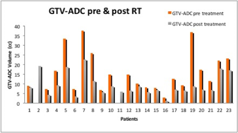
MATERIALS/METHODS: MRI and pathological data collected for 25patients with resectable and borderline resectable PDAC following nCR were retrospectively analyzed. Pre- and post-nCR mean ADC values in the tumors were compared using Wilcoxon matched pairs test. Correlation of pathological treatment response and ADC values was assessed using Pearson's correlation coefficient test and receiver-operating-curve (ROC) analysis.

RESULTS

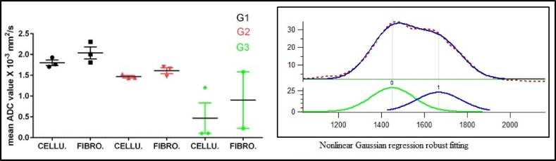
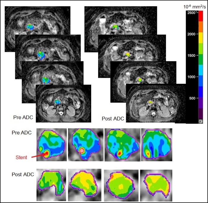
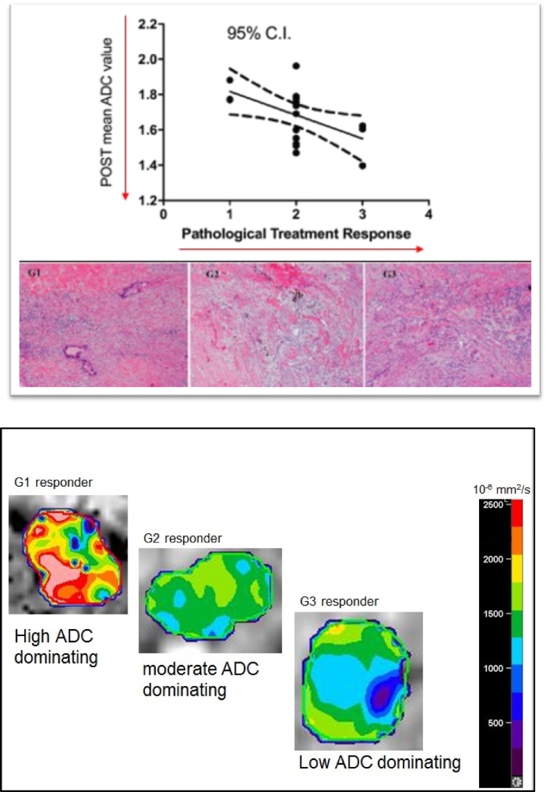
The average mean and standard deviation (SD) of the ADC values for all the patients analyzed were significantly higher in post-nCR (1.667±0.161×10) compared with those prior to nCR (1.395±0.136×10 mm/sec), (P<0.05). The mean ADC values after nCR were significantly correlated with the pathological responses (r=-0.5172); P=0.02. The area under the curve (AUC) of the ADC values for differentiating G1, G2 and G3 pathological responses, using ROC analysis, was found to be 0.6310 and P=0.03.

CONCLUSION

Changes of pre- and post-treatment ADC values significantly correlated with pathological treatment response for PDAC patients treated with chemoradiation therapy, indicating that the ADC could be used to assesstreatment response for PDAC.

摘要

目的

探讨在新辅助放化疗(nCR)后使用表观扩散系数(ADC)评估胰腺导管腺癌(PDAC)病理治疗反应的可行性。

材料/方法:回顾性分析25例nCR后可切除和交界可切除PDAC患者的MRI和病理数据。使用Wilcoxon配对检验比较肿瘤在nCR前后的平均ADC值。使用Pearson相关系数检验和受试者操作特征曲线(ROC)分析评估病理治疗反应与ADC值的相关性。

结果

所有分析患者的ADC值,nCR后的平均均值和标准差(SD)(1.667±0.161×10)显著高于nCR前(1.395±0.136×10 mm²/sec),(P<0.05)。nCR后的平均ADC值与病理反应显著相关(r=-0.5172);P=0.02。使用ROC分析,ADC值区分G1、G2和G3病理反应的曲线下面积(AUC)为0.6310,P=0.03。

结论

放化疗治疗的PDAC患者治疗前后ADC值的变化与病理治疗反应显著相关,表明ADC可用于评估PDAC的治疗反应。

https://cdn.ncbi.nlm.nih.gov/pmc/blobs/d736/5852406/2862255b498b/gr5.jpg
https://cdn.ncbi.nlm.nih.gov/pmc/blobs/d736/5852406/28527384bc63/gr1.jpg
https://cdn.ncbi.nlm.nih.gov/pmc/blobs/d736/5852406/31277b0647f6/gr2.jpg
https://cdn.ncbi.nlm.nih.gov/pmc/blobs/d736/5852406/80a57449692b/gr3.jpg
https://cdn.ncbi.nlm.nih.gov/pmc/blobs/d736/5852406/a995d8dda0ae/gr4.jpg
https://cdn.ncbi.nlm.nih.gov/pmc/blobs/d736/5852406/2862255b498b/gr5.jpg

相似文献

1
Correlation of ADC With Pathological Treatment Response for Radiation Therapy of Pancreatic Cancer.胰腺癌放射治疗中表观扩散系数(ADC)与病理治疗反应的相关性
Transl Oncol. 2018 Apr;11(2):391-398. doi: 10.1016/j.tranon.2018.01.018. Epub 2018 Feb 20.
2
PET-based Treatment Response Assessment for Neoadjuvant Chemoradiation in Pancreatic Adenocarcinoma: An Exploratory Study.基于正电子发射断层扫描的胰腺癌新辅助放化疗治疗反应评估:一项探索性研究
Transl Oncol. 2018 Oct;11(5):1104-1109. doi: 10.1016/j.tranon.2018.06.011. Epub 2018 Jul 17.
3
Endometrial cancer: correlation of apparent diffusion coefficient (ADC) with tumor cellularity and tumor grade.子宫内膜癌:表观扩散系数(ADC)与肿瘤细胞密度及肿瘤分级的相关性
Acta Radiol. 2016 Aug;57(8):1021-8. doi: 10.1177/0284185115612249. Epub 2015 Oct 19.
4
PET/CT in brachytherapy early response evaluation of pancreatic ductal adenocarcinoma xenografts: comparison with apparent diffusion coefficient from diffusion-weighted MR imaging.PET/CT 引导近距离放疗早期评价胰腺导管腺癌移植瘤:与扩散加权磁共振成像表观扩散系数的比较。
Abdom Radiol (NY). 2019 Mar;44(3):950-957. doi: 10.1007/s00261-018-1791-x.
5
Pancreatic neuroendocrine neoplasms at magnetic resonance imaging: comparison between grade 3 and grade 1/2 tumors.磁共振成像下的胰腺神经内分泌肿瘤:3级与1/2级肿瘤的比较
Onco Targets Ther. 2017 Mar 7;10:1465-1474. doi: 10.2147/OTT.S127803. eCollection 2017.
6
Intravoxel incoherent motion magnetic resonance imaging for differentiating metastatic and non-metastatic lymph nodes in pancreatic ductal adenocarcinoma.体素内不相干运动磁共振成像在胰导管腺癌中鉴别转移性和非转移性淋巴结。
Eur Radiol. 2018 Jul;28(7):2781-2789. doi: 10.1007/s00330-017-5259-0. Epub 2018 Feb 5.
7
Comparative analysis of the value of diffusion kurtosis imaging and diffusion-weighted imaging in evaluating the histological features of endometrial cancer.扩散峰度成像与扩散加权成像评估子宫内膜癌组织学特征的价值比较分析。
Cancer Imaging. 2019 Feb 14;19(1):9. doi: 10.1186/s40644-019-0196-6.
8
[Study of diffusion weighted imaging in combination with conventional MRI signs for inflammation activity of chronic hepatitis B].[扩散加权成像联合传统MRI征象对慢性乙型肝炎炎症活动度的研究]
Zhonghua Yi Xue Za Zhi. 2017 May 23;97(19):1484-1490. doi: 10.3760/cma.j.issn.0376-2491.2017.19.012.
9
Diffusion and perfusion MR parameters to assess preoperative short-course radiotherapy response in locally advanced rectal cancer: a comparative explorative study among Standardized Index of Shape by DCE-MRI, intravoxel incoherent motion- and diffusion kurtosis imaging-derived parameters.扩散和灌注磁共振参数评估局部进展期直肠癌术前短程放疗反应:DCE-MRI 标准化形状指数、体素内不相干运动和扩散峰度成像衍生参数的比较探索性研究。
Abdom Radiol (NY). 2019 Nov;44(11):3683-3700. doi: 10.1007/s00261-018-1801-z.
10
Limited Role of the Apparent Diffusion Coefficient (ADC) for Tumor Grade and Overall Survival in Resectable Pancreatic Ductal Adenocarcinoma.表观扩散系数(ADC)在可切除胰腺导管腺癌的肿瘤分级和总生存中的作用有限。
Diagnostics (Basel). 2024 Mar 7;14(6):573. doi: 10.3390/diagnostics14060573.

引用本文的文献

1
Toward diffusion MRI in the diagnosis and treatment of pancreatic cancer.胰腺癌诊断与治疗中的扩散磁共振成像研究进展
Med Oncol. 2025 May 28;42(7):222. doi: 10.1007/s12032-025-02759-5.
2
Values of apparent diffusion coefficient in pancreatic cancer patients receiving neoadjuvant therapy.胰腺癌新辅助治疗患者表观扩散系数的价值。
BMC Cancer. 2024 Sep 18;24(1):1160. doi: 10.1186/s12885-024-12934-y.
3
Prediction of overall survival in patients with locally advanced pancreatic cancer using longitudinal diffusion-weighted MRI.

本文引用的文献

1
Assessment of treatment response during chemoradiation therapy for pancreatic cancer based on quantitative radiomic analysis of daily CTs: An exploratory study.基于每日CT定量影像组学分析的胰腺癌放化疗期间治疗反应评估:一项探索性研究。
PLoS One. 2017 Jun 2;12(6):e0178961. doi: 10.1371/journal.pone.0178961. eCollection 2017.
2
Increased tumour ADC value during chemotherapy predicts improved survival in unresectable pancreatic cancer.化疗期间肿瘤表观扩散系数(ADC)值升高预示不可切除胰腺癌患者的生存期改善。
Eur Radiol. 2016 Jun;26(6):1835-42. doi: 10.1007/s00330-015-3999-2. Epub 2015 Sep 19.
3
Prognostic relevance of apparent diffusion coefficient obtained by diffusion-weighted MRI in pancreatic cancer.
利用纵向扩散加权磁共振成像预测局部晚期胰腺癌患者的总生存期
Front Oncol. 2024 Jul 18;14:1401464. doi: 10.3389/fonc.2024.1401464. eCollection 2024.
4
Diffusion-weighted imaging for determining response to neoadjuvant therapy in pancreatic cancer: a systematic review and meta-analysis.扩散加权成像在评估胰腺癌新辅助治疗反应中的应用:系统评价和荟萃分析。
Eur Radiol. 2024 May;34(5):3238-3248. doi: 10.1007/s00330-023-10381-0. Epub 2023 Nov 1.
5
The Role of MRI Pancreatic Protocol in Assessing Response to Neoadjuvant Therapy for Patients With Borderline Resectable Pancreatic Cancer.磁共振成像胰腺检查方案在评估可切除边缘胰腺癌患者新辅助治疗反应中的作用
Front Oncol. 2022 Jan 13;11:796317. doi: 10.3389/fonc.2021.796317. eCollection 2021.
6
Which Parameter Influences Local Disease-Free Survival after Radiation Therapy Due to Osteolytic Metastasis? A Retrospective Study with Pre- and Post-Radiation Therapy MRI including Diffusion-Weighted Images.哪种参数会影响溶骨性转移放疗后的局部无病生存期?一项包含放疗前后MRI(包括扩散加权成像)的回顾性研究。
J Clin Med. 2021 Dec 25;11(1):106. doi: 10.3390/jcm11010106.
7
Early Response to Chemotherapy in Malignant Pleural Mesothelioma Evaluated Using Diffusion-Weighted Magnetic Resonance Imaging: Initial Observations.使用扩散加权磁共振成像评估恶性胸膜间皮瘤对化疗的早期反应:初步观察
JTO Clin Res Rep. 2021 Nov 2;2(12):100253. doi: 10.1016/j.jtocrr.2021.100253. eCollection 2021 Dec.
8
Longitudinal Monitoring of Simulated Interstitial Fluid Pressure for Pancreatic Ductal Adenocarcinoma Patients Treated with Stereotactic Body Radiotherapy.立体定向体部放射治疗的胰腺导管腺癌患者模拟组织间液压力的纵向监测
Cancers (Basel). 2021 Aug 26;13(17):4319. doi: 10.3390/cancers13174319.
9
A comparative analysis of CT and MRI in differentiating pancreatic cancer from mass pancreatitis.CT与MRI在鉴别胰腺癌与肿块型胰腺炎中的对比分析
Am J Transl Res. 2021 Jun 15;13(6):6431-6438. eCollection 2021.
10
Role of imaging in evaluating the response after neoadjuvant treatment for pancreatic ductal adenocarcinoma.影像学在评估新辅助治疗后胰腺导管腺癌反应中的作用。
World J Gastroenterol. 2021 Jun 14;27(22):3037-3049. doi: 10.3748/wjg.v27.i22.3037.
扩散加权磁共振成像获得的表观扩散系数在胰腺癌中的预后相关性
J Magn Reson Imaging. 2015 Dec;42(6):1532-7. doi: 10.1002/jmri.24939. Epub 2015 May 6.
4
A pilot study of diffusion-weighted MRI in patients undergoing neoadjuvant chemoradiation for pancreatic cancer.胰腺癌新辅助放化疗患者的弥散加权 MRI 初步研究。
Transl Oncol. 2014 Oct 24;7(5):644-9. doi: 10.1016/j.tranon.2014.07.005. eCollection 2014 Oct.
5
Stromal response to Hedgehog signaling restrains pancreatic cancer progression.基质对刺猬信号通路的反应抑制胰腺癌进展。
Proc Natl Acad Sci U S A. 2014 Jul 29;111(30):E3091-100. doi: 10.1073/pnas.1411679111. Epub 2014 Jul 14.
6
The role of diffusion-weighted magnetic resonance imaging in the treatment response evaluation of hepatocellular carcinoma patients treated with radiation therapy.弥散加权磁共振成像在放射治疗肝细胞癌患者治疗反应评估中的作用。
Int J Radiat Oncol Biol Phys. 2014 Jul 15;89(4):814-21. doi: 10.1016/j.ijrobp.2014.03.020.
7
Variability of target and normal structure delineation using multimodality imaging for radiation therapy of pancreatic cancer.使用多模态成像技术对胰腺癌放射治疗进行靶区和正常结构勾画的变异性。
Int J Radiat Oncol Biol Phys. 2014 Jul 1;89(3):633-40. doi: 10.1016/j.ijrobp.2014.02.035. Epub 2014 Apr 20.
8
Predicting and monitoring cancer treatment response with diffusion-weighted MRI.利用扩散加权磁共振成像预测和监测癌症治疗反应。
J Magn Reson Imaging. 2010 Jul;32(1):2-16. doi: 10.1002/jmri.22167.
9
Validation of functional diffusion maps (fDMs) as a biomarker for human glioma cellularity.验证功能扩散映射(fDMs)作为人类脑胶质瘤细胞密度的生物标志物。
J Magn Reson Imaging. 2010 Mar;31(3):538-48. doi: 10.1002/jmri.22068.
10
Cancer. Breaching the cancer fortress.癌症。突破癌症堡垒。
Science. 2009 Jun 12;324(5933):1400-1. doi: 10.1126/science.1175940.