Zhang Yingdi, Ruan Zheng, Shen Minhua, Tan Luanjun, Huang Weiqin, Wang Lei, Huang Yuanliang
Department of Stomatology, East Hospital Affiliated to Tongji University, Shanghai 200120, P.R. China.
Exp Ther Med. 2018 Mar;15(3):2277-2286. doi: 10.3892/etm.2018.5696. Epub 2018 Jan 4.
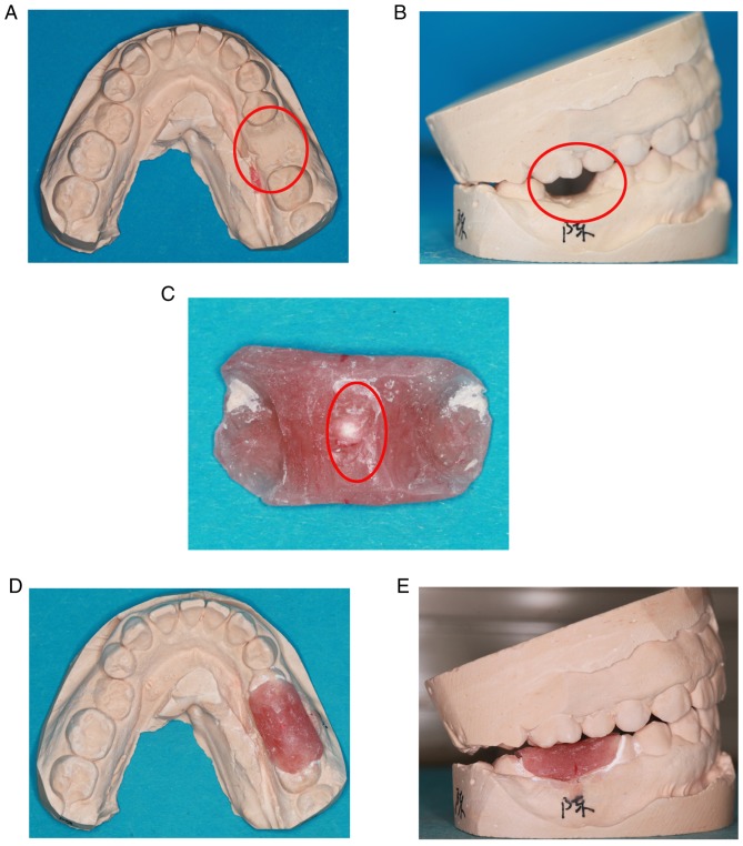
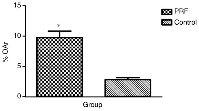
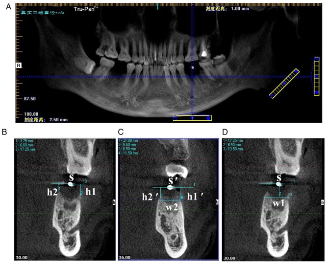
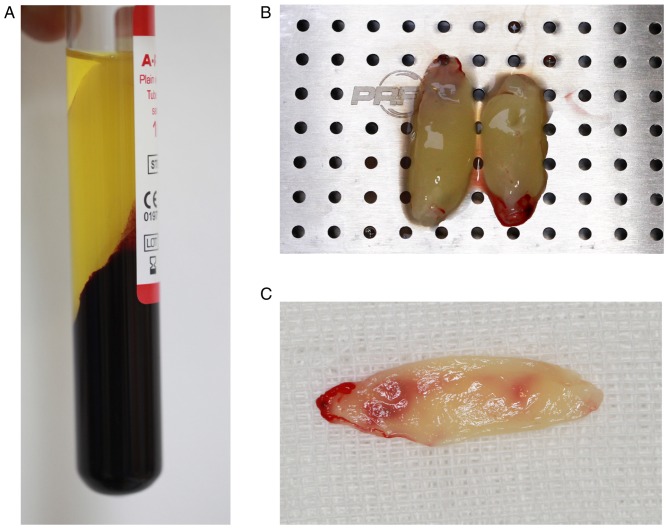
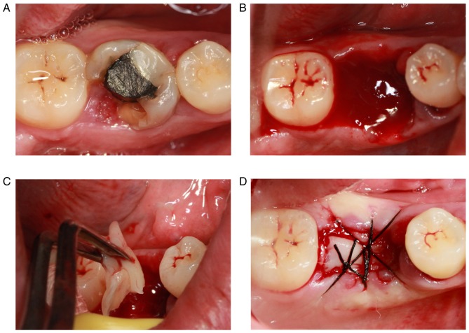
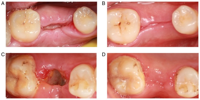
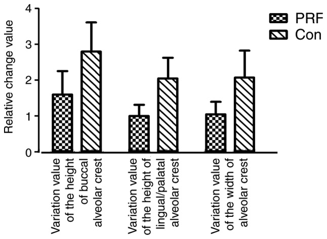
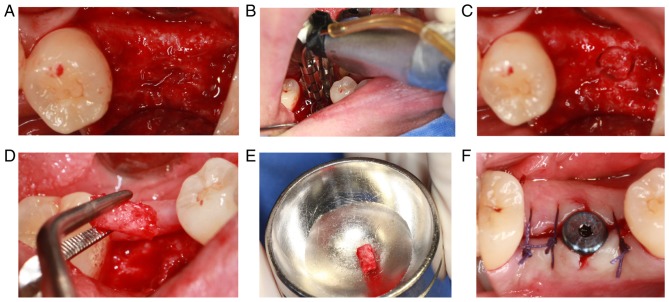
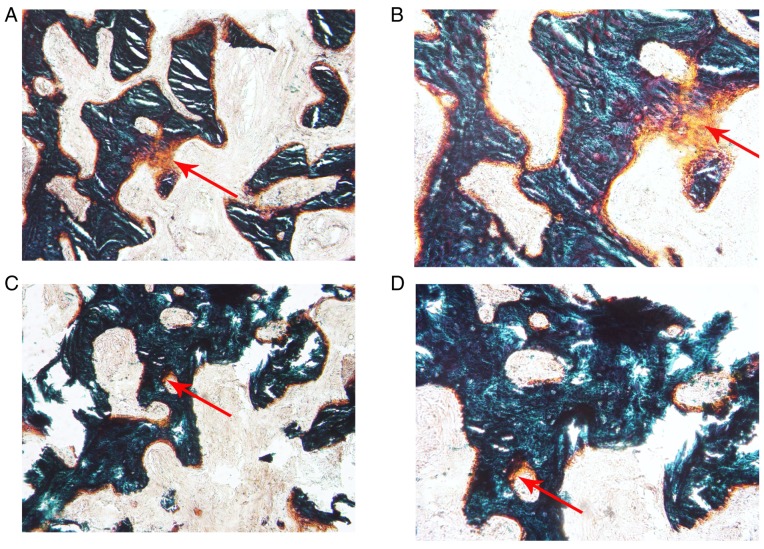
The aim of the present study was to evaluate the clinical efficacy of platelet-rich fibrin (PRF) in preserving the alveolar ridge following human tooth extraction. A total of 28 patients were divided into two groups: The experimental and control groups (n=14 each). Following tooth extraction, the experimental group was implanted with PRF membrane, whereas the control group was not. The gingival healing effect was assessed at 7 days, 1 and 3 months later. Cone-beam computed tomography was performed immediately and at 3 months following tooth extraction. The changes in alveolar ridge height, width, and bone mineral density were compared between the two groups. The alveolar bone was removed using the ring drill during the implant surgery at 3 months following tooth extraction. Histomorphometric evaluation was performed to compare new bone formation between groups. The patients in the experimental group reportedly felt better compared with the patients in the control group. The healing of gingival tissue was better in the experimental group than in the control group. A significantly greater novel bone area was observed in the PRF group compared with the control group (P<0.01). However, no statistically significant differences were observed in the mean value of buccal alveolar ridge height, lingual/palatal alveolar ridge height and alveolar ridge width between the two groups. These results suggested that PRF was advantageous in human alveolar ridge preservation with ease of use and simple handling. Histological analysis of novel bone formation confirmed that PRF increased the quality of the novel bone and enhanced the rate of bone formation, despite the effect of PRF was not significant to reduce alveolar bone resorption in the extraction socket alone.
本研究的目的是评估富血小板纤维蛋白(PRF)在人类拔牙后保存牙槽嵴方面的临床疗效。总共28例患者被分为两组:实验组和对照组(每组n = 14)。拔牙后,实验组植入PRF膜,而对照组未植入。在7天、1个月和3个月后评估牙龈愈合效果。拔牙后立即及3个月时进行锥形束计算机断层扫描。比较两组牙槽嵴高度、宽度和骨密度的变化。在拔牙后3个月进行种植手术时,使用环钻去除牙槽骨。进行组织形态计量学评估以比较组间新骨形成情况。据报道,实验组患者比对照组患者感觉更好。实验组牙龈组织的愈合情况比对照组更好。与对照组相比,PRF组观察到明显更大的新骨面积(P<0.01)。然而,两组之间颊侧牙槽嵴高度、舌侧/腭侧牙槽嵴高度和牙槽嵴宽度的平均值没有观察到统计学上的显著差异。这些结果表明,PRF在人类牙槽嵴保存方面具有优势,使用方便且操作简单。对新骨形成的组织学分析证实,PRF提高了新骨的质量并提高了骨形成速率,尽管PRF单独对减少拔牙窝内牙槽骨吸收的作用不显著。