Yano Mitsutake, Yasuda Masanori, Sakaki Mika, Nagata Koji, Fujino Takashi, Arai Eiichi, Hasebe Takahiro, Miyazawa Masaki, Miyazawa Mariko, Ogane Naoki, Hasegawa Kosei, Narahara Hisashi
Department of Pathology, Saitama Medical University International Medical Center, Saitama 350-1298, Japan.
Department of Obstetrics and Gynecology, Oita University Faculty of Medicine, Oita 879-5593, Japan.
Oncol Lett. 2018 Mar;15(3):3524-3531. doi: 10.3892/ol.2018.7726. Epub 2018 Jan 4.
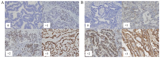
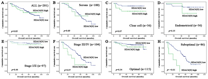
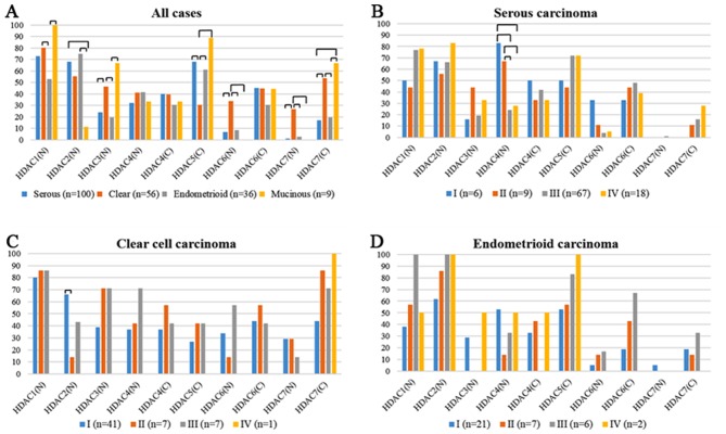
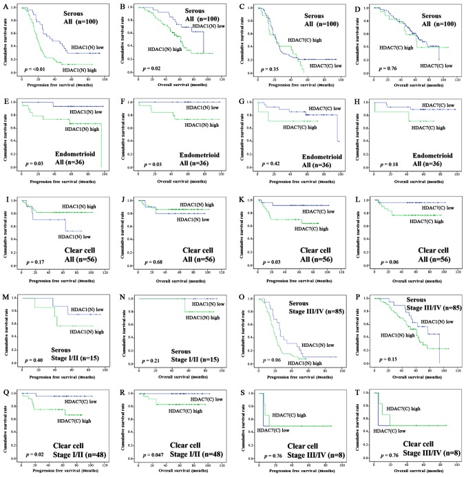
Histone deacetylase (HDAC) inhibitor is known to have a cytotoxic effect on ovarian cancer cell lines. The present study analyzed the association between immunohistochemical HDAC expression and clinicopathological findings, in particular, the association with histological type and effect of chemotherapy. The histology of the 201 ovarian cancers addressed was as follows: Serous carcinoma (SEC), 100 cases; clear cell carcinoma (CCC), 56 cases; endometrioid carcinoma (EMC), 36 cases; and mucinous carcinoma (MUC), 9 cases. Immunohistochemical analyses of HDACs 1, 2, 3, 4, 5, 6 and 7 expression levels were performed using tissue microarrays, composed of 201 primary tumors and 38 tumors following chemotherapy. Overexpression of HDAC1 was detected in the nucleus of all cases with MUC, followed by CCC (80%), SEC (73%), and EMC (53%). CCC specifically demonstrated HDAC7 expression in both the nucleus (27%) and the cytoplasm (54%), and HDAC6 expression in the nucleus (34%). The comparison between prior to and following chemotherapy revealed a nuclear expression increase in HDAC1 (76% vs. 92%; P=0.03) and HDAC7 (0.0 vs. 16%; P=0.01), and cytoplasmic expression increase in HDAC6 (40 vs. 74%; P=<0.01) and HDAC7 (16 vs. 66%; P=<0.01). HDAC1 nuclear expression adversely affected overall survival in SEC (P=0.02) and EMC (P=0.03), and HDAC7 cytoplasmic expression in CCC was associated with a poor prognosis (P=0.06). In multivariate analysis, HDAC6 nuclear expression was determined as a poor prognostic factor (hazard ratio=3.51; 95% confidence interval, 1.49 to 8.27, P=<0.01). In the subgroup analysis, HDAC6 nuclear expression was associated with a poor prognosis in CCC (P=0.07), International Federation of Obstetrics and Gynecology stage III/IV (P=0.07), and suboptimal surgery (P=<0.01). In conclusion, HDACs may be associated with the prognosis of ovarian cancers, depending on the histological subtypes, and upregulated following chemotherapy. HDAC1, 6 and 7 may therefor act as promising therapeutic targets in the future.
已知组蛋白去乙酰化酶(HDAC)抑制剂对卵巢癌细胞系具有细胞毒性作用。本研究分析了免疫组化HDAC表达与临床病理结果之间的关联,特别是与组织学类型及化疗效果的关联。所研究的201例卵巢癌的组织学类型如下:浆液性癌(SEC)100例;透明细胞癌(CCC)56例;子宫内膜样癌(EMC)36例;黏液性癌(MUC)9例。使用由201个原发性肿瘤和38个化疗后肿瘤组成的组织芯片,对HDAC1、2、3、4、5、6和7的表达水平进行免疫组化分析。在所有MUC病例的细胞核中均检测到HDAC1过表达,其次是CCC(80%)、SEC(73%)和EMC(53%)。CCC在细胞核(27%)和细胞质(54%)中均特异性显示HDAC7表达,在细胞核中显示HDAC6表达(34%)。化疗前后的比较显示,HDAC1的核表达增加(76%对92%;P=0.03)和HDAC7的核表达增加(0.0对16%;P=0.01),HDAC6的细胞质表达增加(40对74%;P<0.01)和HDAC7的细胞质表达增加(16对66%;P<0.01)。HDAC1核表达对SEC(P=0.02)和EMC(P=0.03)的总生存期有不利影响,CCC中HDAC7细胞质表达与预后不良相关(P=0.06)。在多因素分析中,HDAC6核表达被确定为不良预后因素(风险比=3.51;95%置信区间,1.49至8.27,P<0.01)。在亚组分析中,HDAC6核表达与CCC(P=0.07)、国际妇产科联合会III/IV期(P=0.07)和手术不充分(P<0.01)的预后不良相关。总之,HDAC可能与卵巢癌的预后相关,取决于组织学亚型,且化疗后上调。因此,HDAC1、6和7可能在未来成为有前景的治疗靶点。