Fujimoto Yuji, Tomimaru Yoshito, Hatano Hisanori, Noguchi Kozo, Nagase Hirotsugu, Hamabe Atsushi, Hirota Masashi, Oshima Kazuteru, Tanida Tsukasa, Morita Shunji, Imamura Hiroshi, Iwazawa Takashi, Akagi Kenzo, Dono Keizo
Department of Surgery, Toyonaka Municipal Hospital, Toyonaka, Osaka, Japan.
Department of Surgery, Rinku General Medical Center, Izumisano, Osaka, Japan.
Am J Case Rep. 2018 Feb 20;19:187-193. doi: 10.12659/ajcr.907273.
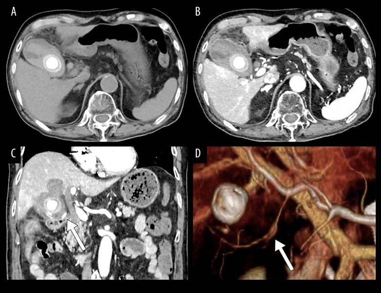
BACKGROUND Cystic artery pseudoaneurysm is rare, and some cases are associated with inflammation of the gallbladder. There is limited information regarding this condition, and the clinical features remain unclear. This report is a case of ruptured cystic artery pseudoaneurysm diagnosed by computed tomography (CT) imaging and treated with urgent cholecystectomy and is supported by a literature review of previous cases. CASE REPORT A 90-year-old man, who had developed acute cholecystitis due to a gallstone one month previously, was referred to our hospital. He developed fever and epigastric pain while waiting for a scheduled elective cholecystectomy. Laboratory investigations showed elevated markers of inflammation and elevated hepatobiliary enzyme levels. Computed tomography (CT) imaging showed cholecystitis and pseudoaneurysm of the cystic artery. The pseudoaneurysm had ruptured and was accompanied by the formation of a hematoma within the gallbladder that involved the liver bed. Having made the preoperative diagnosis, an urgent open laparotomy was performed, during which the gallbladder was found to have perforated. The hematoma penetrated into the liver bed. Cholecystectomy was performed, and the pseudoaneurysm of the cystic artery was extirpated. There were no serious postoperative complications. A literature review identified 50 previously reported case of cystic artery pseudoaneurysm. CONCLUSIONS A case of ruptured cystic artery pseudoaneurysm, successfully treated with urgent cholecystectomy is reported, supported by a literature review of previous cases and characterization of the clinical features of this rare condition.
胆囊动脉假性动脉瘤较为罕见,部分病例与胆囊炎相关。关于这种情况的信息有限,临床特征仍不明确。本报告为一例经计算机断层扫描(CT)成像诊断为胆囊动脉假性动脉瘤破裂,并接受紧急胆囊切除术治疗的病例,并通过对既往病例的文献回顾提供支持。
一名90岁男性,1个月前因胆结石引发急性胆囊炎,转诊至我院。在等待择期胆囊切除术期间,他出现发热和上腹部疼痛。实验室检查显示炎症指标升高和肝胆酶水平升高。计算机断层扫描(CT)成像显示胆囊炎和胆囊动脉假性动脉瘤。假性动脉瘤已破裂,并伴有胆囊内血肿形成,累及肝床。在做出术前诊断后,进行了紧急剖腹手术,术中发现胆囊已穿孔。血肿穿透至肝床。进行了胆囊切除术,并切除了胆囊动脉假性动脉瘤。术后无严重并发症。文献回顾确定了50例先前报道的胆囊动脉假性动脉瘤病例。
报告了一例经紧急胆囊切除术成功治疗的胆囊动脉假性动脉瘤破裂病例,并通过对既往病例的文献回顾以及对这种罕见疾病临床特征的描述提供支持。