• 文献检索
  • 文档翻译
  • 深度研究
  • 学术资讯
  • Suppr Zotero 插件Zotero 插件
  • 邀请有礼
  • 套餐&价格
  • 历史记录
应用&插件
Suppr Zotero 插件Zotero 插件浏览器插件Mac 客户端Windows 客户端微信小程序
定价
高级版会员购买积分包购买API积分包
服务
文献检索文档翻译深度研究API 文档MCP 服务
关于我们
关于 Suppr公司介绍联系我们用户协议隐私条款
关注我们

Suppr 超能文献

核心技术专利:CN118964589B侵权必究
粤ICP备2023148730 号-1Suppr @ 2026

文献检索

告别复杂PubMed语法,用中文像聊天一样搜索,搜遍4000万医学文献。AI智能推荐,让科研检索更轻松。

立即免费搜索

文件翻译

保留排版,准确专业,支持PDF/Word/PPT等文件格式,支持 12+语言互译。

免费翻译文档

深度研究

AI帮你快速写综述,25分钟生成高质量综述,智能提取关键信息,辅助科研写作。

立即免费体验

肿瘤起始细胞诱导 Cxcr4 介导的促肿瘤巨噬细胞浸润大脑。

Tumor initiating cells induce Cxcr4-mediated infiltration of pro-tumoral macrophages into the brain.

机构信息

Centre for Discovery Brain Sciences, University of Edinburgh, Edinburgh, United Kingdom.

Centre for Integrative Biology (CIBIO), University of Trento, Trento, Italy.

出版信息

Elife. 2018 Feb 21;7:e31918. doi: 10.7554/eLife.31918.

DOI:10.7554/eLife.31918
PMID:29465400
原文链接:https://pmc.ncbi.nlm.nih.gov/articles/PMC5821457/
Abstract

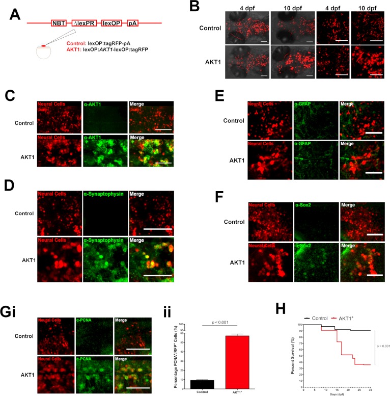
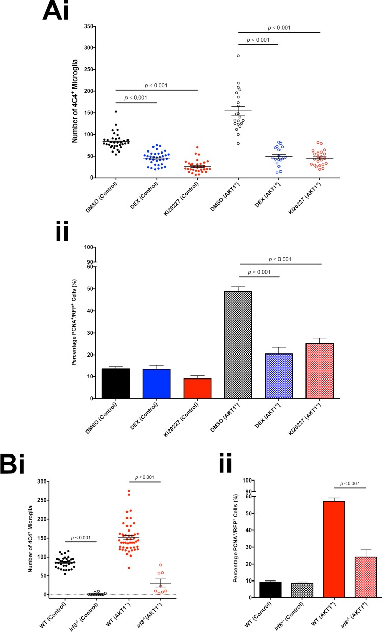
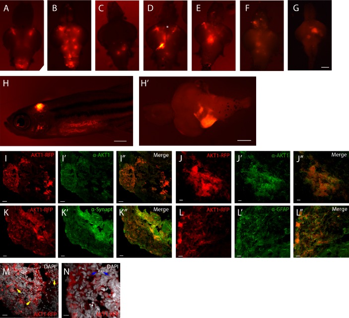
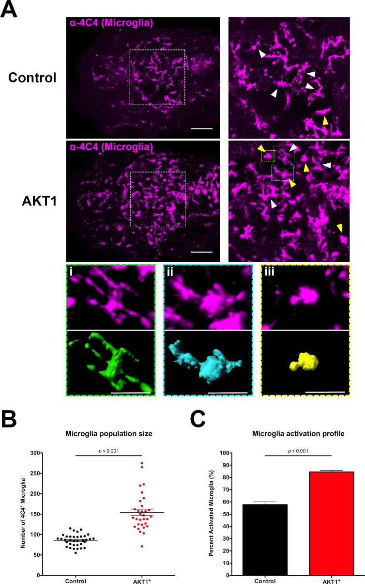
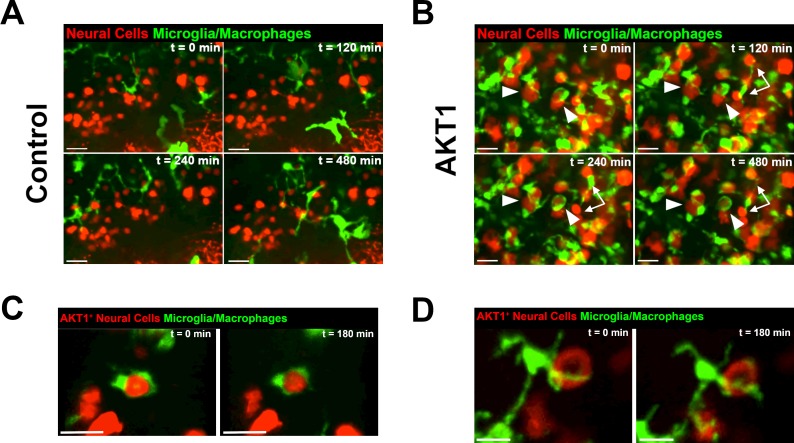
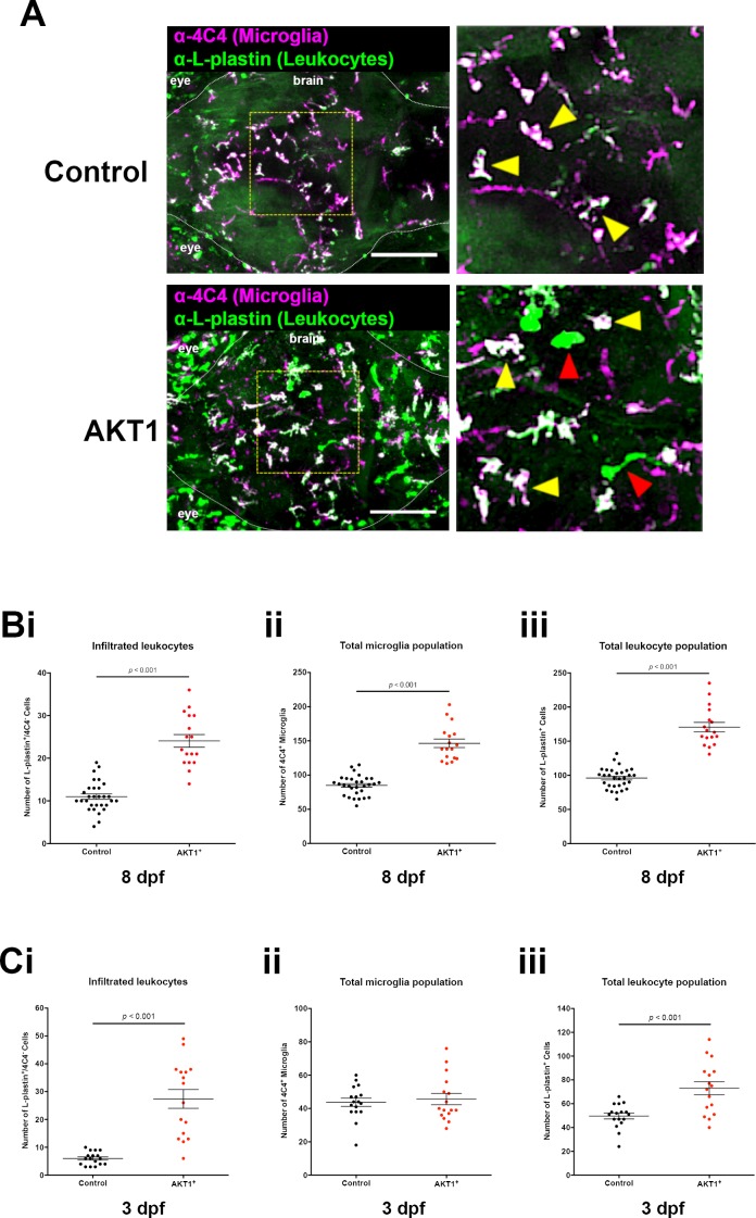
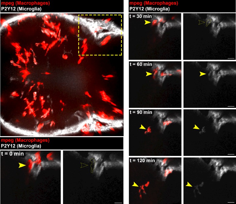
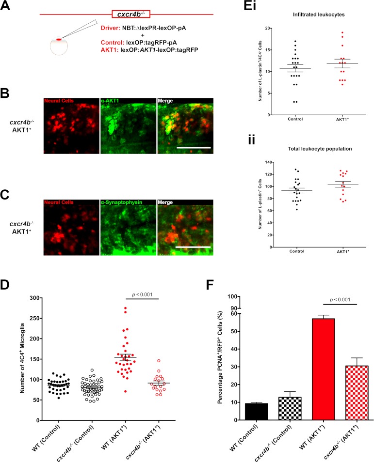
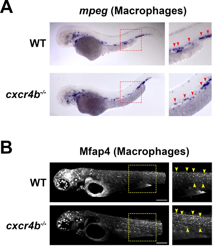
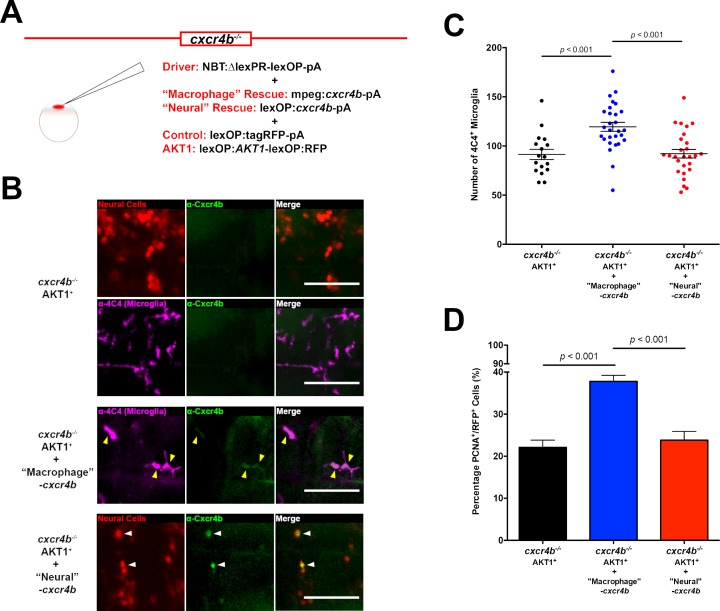
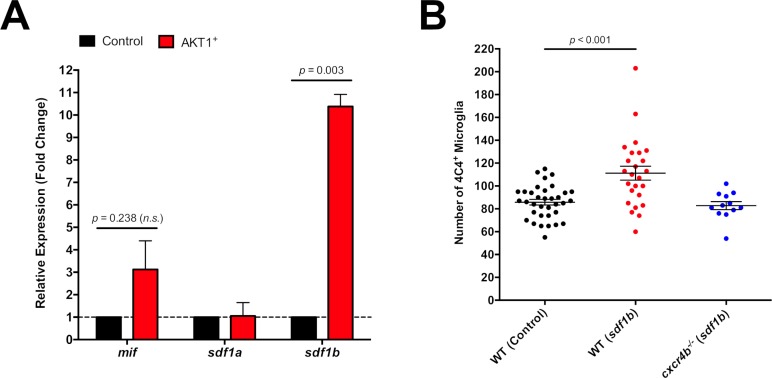
It is now clear that microglia and macrophages are present in brain tumors, but whether or how they affect initiation and development of tumors is not known. Exploiting the advantages of the zebrafish () model, we showed that macrophages and microglia respond immediately upon oncogene activation in the brain. Overexpression of human AKT1 within neural cells of larval zebrafish led to a significant increase in the macrophage and microglia populations. By using a combination of transgenic and mutant zebrafish lines, we showed that this increase was caused by the infiltration of peripheral macrophages into the brain mediated via Sdf1b-Cxcr4b signaling. Intriguingly, confocal live imaging reveals highly dynamic interactions between macrophages/microglia and pre-neoplastic cells, which do not result in phagocytosis of pre-neoplastic cells. Finally, depletion of macrophages and microglia resulted in a significant reduction of oncogenic cell proliferation. Thus, macrophages and microglia show tumor promoting functions already during the earliest stages of the developing tumor microenvironment.

摘要

现在已经清楚的是,小胶质细胞和巨噬细胞存在于脑肿瘤中,但它们是否以及如何影响肿瘤的发生和发展尚不清楚。利用斑马鱼()模型的优势,我们发现巨噬细胞和小胶质细胞在大脑中的癌基因激活后立即作出反应。在幼鱼斑马鱼的神经细胞中过表达人 AKT1,会导致巨噬细胞和小胶质细胞群体显著增加。通过使用转基因和突变斑马鱼系的组合,我们表明这种增加是由 Sdf1b-Cxcr4b 信号介导的外周巨噬细胞浸润到大脑中引起的。有趣的是,共聚焦活细胞成像揭示了巨噬细胞/小胶质细胞与前瘤细胞之间高度动态的相互作用,但不会导致前瘤细胞的吞噬。最后,耗尽巨噬细胞和小胶质细胞会导致致癌细胞增殖的显著减少。因此,巨噬细胞和小胶质细胞在肿瘤微环境发育的最早阶段就表现出促进肿瘤的功能。

https://cdn.ncbi.nlm.nih.gov/pmc/blobs/7997/5821457/47027b3e7be9/elife-31918-fig8.jpg
https://cdn.ncbi.nlm.nih.gov/pmc/blobs/7997/5821457/ee787ef3c497/elife-31918-fig1.jpg
https://cdn.ncbi.nlm.nih.gov/pmc/blobs/7997/5821457/ee7a9360bd9b/elife-31918-fig1-figsupp1.jpg
https://cdn.ncbi.nlm.nih.gov/pmc/blobs/7997/5821457/2991ffc99322/elife-31918-fig2.jpg
https://cdn.ncbi.nlm.nih.gov/pmc/blobs/7997/5821457/52345a7bdf4c/elife-31918-fig3.jpg
https://cdn.ncbi.nlm.nih.gov/pmc/blobs/7997/5821457/eff23c52f96d/elife-31918-fig4.jpg
https://cdn.ncbi.nlm.nih.gov/pmc/blobs/7997/5821457/209bdf616570/elife-31918-fig4-figsupp1.jpg
https://cdn.ncbi.nlm.nih.gov/pmc/blobs/7997/5821457/19fe01ac4509/elife-31918-fig5.jpg
https://cdn.ncbi.nlm.nih.gov/pmc/blobs/7997/5821457/f9074f707e20/elife-31918-fig5-figsupp1.jpg
https://cdn.ncbi.nlm.nih.gov/pmc/blobs/7997/5821457/93aa03408b4e/elife-31918-fig6.jpg
https://cdn.ncbi.nlm.nih.gov/pmc/blobs/7997/5821457/8f29a503a332/elife-31918-fig7.jpg
https://cdn.ncbi.nlm.nih.gov/pmc/blobs/7997/5821457/47027b3e7be9/elife-31918-fig8.jpg

相似文献

1
Tumor initiating cells induce Cxcr4-mediated infiltration of pro-tumoral macrophages into the brain.肿瘤起始细胞诱导 Cxcr4 介导的促肿瘤巨噬细胞浸润大脑。
Elife. 2018 Feb 21;7:e31918. doi: 10.7554/eLife.31918.
2
Involvement of Cxcl12a/Cxcr4b Chemokine System in Mediating the Stimulatory Effect of Embryonic Ethanol Exposure on Neuronal Density in Zebrafish Hypothalamus.Cxcl12a/Cxcr4b 趋化因子系统在介导胚胎乙醇暴露对斑马鱼下丘脑神经元密度的刺激作用中的作用。
Alcohol Clin Exp Res. 2020 Dec;44(12):2519-2535. doi: 10.1111/acer.14482. Epub 2020 Nov 16.
3
A syngeneic spontaneous zebrafish model of -deficient, EGFR, and PI3KCA-driven glioblastoma reveals inhibitory roles for inflammation during tumor initiation and relapse in vivo.-/-/- 缺陷型、EGFR 和 PI3KCA 驱动的胶质母细胞瘤的同基因自发斑马鱼模型揭示了体内肿瘤起始和复发过程中炎症的抑制作用。
Elife. 2024 Jul 25;13:RP93077. doi: 10.7554/eLife.93077.
4
CXCR4 signaling regulates metastatic onset by controlling neutrophil motility and response to malignant cells.CXCR4 信号通过控制中性粒细胞的运动和对恶性细胞的反应来调节转移的起始。
Sci Rep. 2019 Feb 20;9(1):2399. doi: 10.1038/s41598-019-38643-2.
5
PTEN loss mediated Akt activation promotes prostate tumor growth and metastasis via CXCL12/CXCR4 signaling.PTEN 缺失介导的 Akt 激活通过 CXCL12/CXCR4 信号促进前列腺肿瘤生长和转移。
Mol Cancer. 2013 Jul 31;12(1):85. doi: 10.1186/1476-4598-12-85.
6
Notch1 signaling pathway promotes invasion, self-renewal and growth of glioma initiating cells via modulating chemokine system CXCL12/CXCR4.Notch1 信号通路通过调节趋化因子系统 CXCL12/CXCR4 促进胶质瘤起始细胞的侵袭、自我更新和生长。
J Exp Clin Cancer Res. 2019 Aug 5;38(1):339. doi: 10.1186/s13046-019-1319-4.
7
Tetraspanin Cd9b and Cxcl12a/Cxcr4b have a synergistic effect on the control of collective cell migration.四跨膜蛋白 Cd9b 和 Cxcl12a/Cxcr4b 对细胞集体迁移的控制具有协同作用。
PLoS One. 2021 Nov 30;16(11):e0260372. doi: 10.1371/journal.pone.0260372. eCollection 2021.
8
CXCR4 mammary gland macrophageal niche promotes tumor initiating cell activity and immune suppression during tumorigenesis.CXCR4乳腺巨噬细胞生态位在肿瘤发生过程中促进肿瘤起始细胞活性和免疫抑制。
Nat Commun. 2025 May 25;16(1):4854. doi: 10.1038/s41467-025-59972-z.
9
Programmed Cell Death 10 Mediated CXCL2-CXCR2 Signaling in Regulating Tumor-Associated Microglia/Macrophages Recruitment in Glioblastoma.程序性细胞死亡分子 10 介导的 CXCL2-CXCR2 信号在调节胶质母细胞瘤中肿瘤相关小胶质细胞/巨噬细胞募集中的作用。
Front Immunol. 2021 May 24;12:637053. doi: 10.3389/fimmu.2021.637053. eCollection 2021.
10
Brain tumours repurpose endogenous neuron to microglia signalling mechanisms to promote their own proliferation.脑肿瘤将内源性神经元重编程为小胶质细胞信号机制,以促进自身增殖。
Elife. 2019 Jul 17;8:e46912. doi: 10.7554/eLife.46912.

引用本文的文献

1
Calciumnetexplorer: an R package for network analysis of calcium imaging data.钙网络浏览器:一个用于钙成像数据网络分析的R软件包。
BMC Bioinformatics. 2025 Aug 26;26(1):220. doi: 10.1186/s12859-025-06206-0.
2
Melanocyte-dependent macrophage redistribution enhances skin immunity upon acute stress.黑素细胞依赖性巨噬细胞重新分布可增强急性应激时的皮肤免疫力。
Proc Natl Acad Sci U S A. 2025 Aug 26;122(34):e2511358122. doi: 10.1073/pnas.2511358122. Epub 2025 Aug 19.
3
CXCR4 mammary gland macrophageal niche promotes tumor initiating cell activity and immune suppression during tumorigenesis.

本文引用的文献

1
The chemokine receptor CXCR4 promotes granuloma formation by sustaining a mycobacteria-induced angiogenesis programme.趋化因子受体 CXCR4 通过维持分枝杆菌诱导的血管生成程序促进肉芽肿形成。
Sci Rep. 2017 Mar 23;7:45061. doi: 10.1038/srep45061.
2
Cellular and Molecular Identity of Tumor-Associated Macrophages in Glioblastoma.胶质母细胞瘤中肿瘤相关巨噬细胞的细胞和分子特征
Cancer Res. 2017 May 1;77(9):2266-2278. doi: 10.1158/0008-5472.CAN-16-2310. Epub 2017 Feb 24.
3
Involvement of the phosphoinositide 3-kinase/Akt signaling pathway in bone morphogenetic protein 9-stimulated osteogenic differentiation and stromal cell-derived factor 1 production in human periodontal ligament fibroblasts.
CXCR4乳腺巨噬细胞生态位在肿瘤发生过程中促进肿瘤起始细胞活性和免疫抑制。
Nat Commun. 2025 May 25;16(1):4854. doi: 10.1038/s41467-025-59972-z.
4
Elf1 Deficiency Impairs Macrophage Development in Zebrafish Model Organism.Elf1基因缺陷损害斑马鱼模式生物中的巨噬细胞发育。
Int J Mol Sci. 2025 Mar 12;26(6):2537. doi: 10.3390/ijms26062537.
5
Divergent Crosstalk Between Microglia and T Cells in Brain Cancers: Implications for Novel Therapeutic Strategies.脑癌中微胶质细胞与T细胞之间的不同串扰:对新型治疗策略的启示
Biomedicines. 2025 Jan 16;13(1):216. doi: 10.3390/biomedicines13010216.
6
Stat3 mediates Fyn kinase-driven dopaminergic neurodegeneration and microglia activation.信号转导和转录激活因子3介导Fyn激酶驱动的多巴胺能神经变性和小胶质细胞激活。
Dis Model Mech. 2024 Dec 1;17(12). doi: 10.1242/dmm.052011. Epub 2024 Dec 6.
7
Correction: Microglia are essential for tissue contraction in wound closure after brain injury in zebrafish larvae.更正:在斑马鱼幼体脑损伤后的伤口闭合过程中,小胶质细胞对于组织收缩至关重要。
Life Sci Alliance. 2024 Nov 25;8(2). doi: 10.26508/lsa.202403129. Print 2025 Feb.
8
Profiling the cancer-prone microenvironment in a zebrafish model for MPNST.在斑马鱼模型中分析MPNST的癌症易感性微环境。
Oncogene. 2025 Feb;44(3):179-191. doi: 10.1038/s41388-024-03210-1. Epub 2024 Nov 7.
9
Therapeutic approaches to modulate the immune microenvironment in gliomas.调节神经胶质瘤免疫微环境的治疗方法。
NPJ Precis Oncol. 2024 Oct 23;8(1):241. doi: 10.1038/s41698-024-00717-4.
10
Microglia are essential for tissue contraction in wound closure after brain injury in zebrafish larvae.小胶质细胞对于斑马鱼幼体脑损伤后伤口闭合过程中的组织收缩至关重要。
Life Sci Alliance. 2024 Oct 17;8(1). doi: 10.26508/lsa.202403052. Print 2025 Jan.
磷酸肌醇3-激酶/蛋白激酶B信号通路参与骨形态发生蛋白9刺激人牙周膜成纤维细胞的成骨分化及基质细胞衍生因子1的产生。
Eur J Oral Sci. 2017 Apr;125(2):119-126. doi: 10.1111/eos.12336. Epub 2017 Feb 13.
4
Investigating microglia-brain tumor cell interactions in vivo in the larval zebrafish brain.研究斑马鱼幼体大脑中体内小胶质细胞与脑肿瘤细胞的相互作用。
Methods Cell Biol. 2017;138:593-626. doi: 10.1016/bs.mcb.2016.10.001. Epub 2016 Oct 31.
5
A novel brain tumour model in zebrafish reveals the role of YAP activation in MAPK- and PI3K-induced malignant growth.一种新型斑马鱼脑肿瘤模型揭示了YAP激活在MAPK和PI3K诱导的恶性生长中的作用。
Dis Model Mech. 2017 Jan 1;10(1):15-28. doi: 10.1242/dmm.026500. Epub 2016 Nov 24.
6
Macrophage Ontogeny Underlies Differences in Tumor-Specific Education in Brain Malignancies.巨噬细胞个体发生是脑恶性肿瘤中肿瘤特异性驯化差异的基础。
Cell Rep. 2016 Nov 22;17(9):2445-2459. doi: 10.1016/j.celrep.2016.10.052. Epub 2016 Nov 10.
7
A Zebrafish Live Imaging Model Reveals Differential Responses of Microglia Toward Glioblastoma Cells In Vivo.斑马鱼活体成像模型揭示了小胶质细胞在体内对胶质母细胞瘤细胞的不同反应。
Zebrafish. 2016 Dec;13(6):523-534. doi: 10.1089/zeb.2016.1339. Epub 2016 Oct 25.
8
Identification of a conserved and acute neurodegeneration-specific microglial transcriptome in the zebrafish.斑马鱼中保守且急性神经变性特异性小胶质细胞转录组的鉴定。
Glia. 2017 Jan;65(1):138-149. doi: 10.1002/glia.23083. Epub 2016 Oct 19.
9
Developmental Apoptosis Mediates Entry and Positioning of Microglia in the Zebrafish Brain.发育性细胞凋亡介导小胶质细胞进入斑马鱼大脑并定位。
Cell Rep. 2016 Jul 26;16(4):897-906. doi: 10.1016/j.celrep.2016.06.033. Epub 2016 Jul 14.
10
Microglia Colonization of Developing Zebrafish Midbrain Is Promoted by Apoptotic Neuron and Lysophosphatidylcholine.小胶质细胞对发育中斑马鱼中脑的定植受凋亡神经元和溶血磷脂酰胆碱的促进。
Dev Cell. 2016 Jul 25;38(2):214-22. doi: 10.1016/j.devcel.2016.06.018. Epub 2016 Jul 14.