Tissue Engineering Laboratory, Skeletal Biology and Engineering Research Center, KU Leuven, Campus Gasthuisberg O&N 1, Herestraat 49, bus 813, 3000, Leuven, Belgium.
Prometheus, Division of Skeletal Tissue Engineering, KU Leuven, O&N 1, Herestraat 49, bus 813, 3000, Leuven, Belgium.
Stem Cell Res Ther. 2018 Feb 21;9(1):42. doi: 10.1186/s13287-018-0787-3.
Chondrogenic mesenchymal stem cells (MSCs) have not yet been used to address the clinical demands of large osteochondral joint surface defects. In this study, self-assembling tissue intermediates (TIs) derived from human periosteum-derived stem/progenitor cells (hPDCs) were generated and validated for stable cartilage formation in vivo using two different animal models.
hPDCs were aggregated and cultured in the presence of a novel growth factor (GF) cocktail comprising of transforming growth factor (TGF)-β1, bone morphogenetic protein (BMP)2, growth differentiation factor (GDF)5, BMP6, and fibroblast growth factor (FGF)2. Quantitative polymerase chain reaction (PCR) and immunohistochemistry were used to study in vitro differentiation. Aggregates were then implanted ectopically in nude mice and orthotopically in critical-size osteochondral defects in nude rats and evaluated by microcomputed tomography (µCT) and immunohistochemistry.
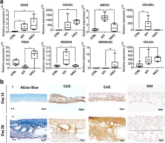
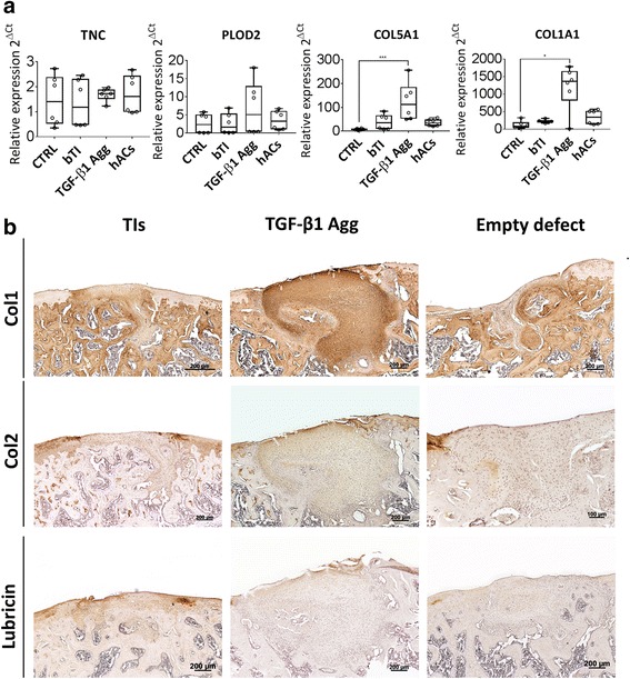
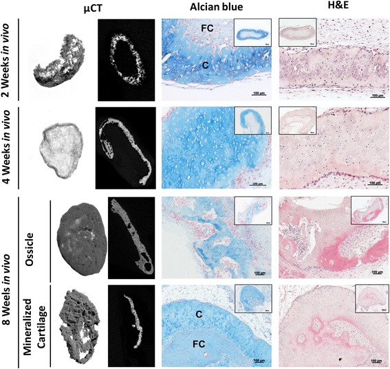
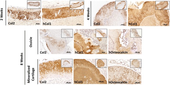
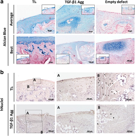
Gene expression analysis after 28 days of in vitro culture revealed the expression of early and late chondrogenic markers and a significant upregulation of NOGGIN as compared to human articular chondrocytes (hACs). Histological examination revealed a bilayered structure comprising of chondrocytes at different stages of maturity. Ectopically, TIs generated both bone and mineralized cartilage at 8 weeks after implantation. Osteochondral defects treated with TIs displayed glycosaminoglycan (GAG) production, type-II collagen, and lubricin expression. Immunostaining for human nuclei protein suggested that hPDCs contributed to both subchondral bone and articular cartilage repair.
Our data indicate that in vitro derived osteochondral-like tissues can be generated from hPDCs, which are capable of producing bone and cartilage ectopically and behave orthotopically as osteochondral units.
软骨形成间充质干细胞(MSCs)尚未用于解决大的骨软骨关节面缺损的临床需求。在这项研究中,使用两种不同的动物模型,从人骨膜源性干细胞/祖细胞(hPDC)中生成和验证了自组装组织中间物(TIs),以实现体内稳定的软骨形成。
在含有转化生长因子(TGF)-β1、骨形态发生蛋白(BMP)2、生长分化因子(GDF)5、BMP6 和成纤维细胞生长因子(FGF)2 的新型生长因子(GF)鸡尾酒存在下,聚集和培养 hPDC。使用定量聚合酶链反应(PCR)和免疫组织化学研究体外分化。然后将聚集物异位植入裸鼠和裸鼠的临界大小骨软骨缺损中,并通过微计算机断层扫描(µCT)和免疫组织化学进行评估。
体外培养 28 天后的基因表达分析显示,与人类关节软骨细胞(hAC)相比,早期和晚期软骨形成标志物的表达以及 NOGGIN 的显著上调。组织学检查显示出具有不同成熟阶段的软骨细胞的双层结构。异位,TIs 在植入后 8 周内产生了骨和矿化软骨。用 TIs 治疗的骨软骨缺损显示糖胺聚糖(GAG)产生、II 型胶原和润滑素表达。人核蛋白免疫染色表明 hPDC 有助于软骨下骨和关节软骨的修复。
我们的数据表明,体外衍生的骨软骨样组织可以从 hPDC 中生成,它们能够异位产生骨和软骨,并作为骨软骨单位发挥同源作用。