• 文献检索
  • 文档翻译
  • 深度研究
  • 学术资讯
  • Suppr Zotero 插件Zotero 插件
  • 邀请有礼
  • 套餐&价格
  • 历史记录
应用&插件
Suppr Zotero 插件Zotero 插件浏览器插件Mac 客户端Windows 客户端微信小程序
定价
高级版会员购买积分包购买API积分包
服务
文献检索文档翻译深度研究API 文档MCP 服务
关于我们
关于 Suppr公司介绍联系我们用户协议隐私条款
关注我们

Suppr 超能文献

核心技术专利:CN118964589B侵权必究
粤ICP备2023148730 号-1Suppr @ 2026

文献检索

告别复杂PubMed语法,用中文像聊天一样搜索,搜遍4000万医学文献。AI智能推荐,让科研检索更轻松。

立即免费搜索

文件翻译

保留排版,准确专业,支持PDF/Word/PPT等文件格式,支持 12+语言互译。

免费翻译文档

深度研究

AI帮你快速写综述,25分钟生成高质量综述,智能提取关键信息,辅助科研写作。

立即免费体验

采用新型静电纺丝方法制备具有径向排列图案的透明半球形 3D 纳米纤维支架。

Fabrication of transparent hemispherical 3D nanofibrous scaffolds with radially aligned patterns via a novel electrospinning method.

机构信息

Department of Bionanosystem Engineering, Graduate School, Chonbuk National University, Jeonju, 561-756, Republic of Korea.

Division of Mechanical Design Engineering, College of Engineering, Chonbuk National University, Jeonju, 561-756, Republic of Korea.

出版信息

Sci Rep. 2018 Feb 21;8(1):3424. doi: 10.1038/s41598-018-21618-0.

DOI:10.1038/s41598-018-21618-0
PMID:29467436
原文链接:https://pmc.ncbi.nlm.nih.gov/articles/PMC5821851/
Abstract

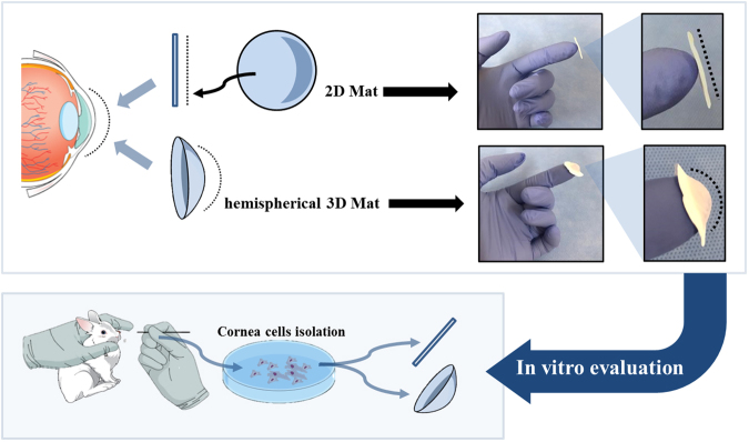
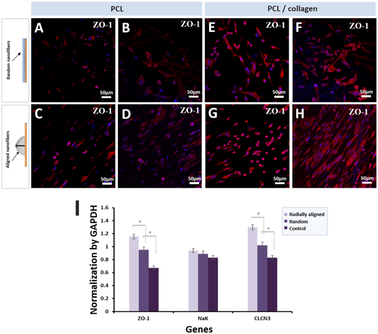
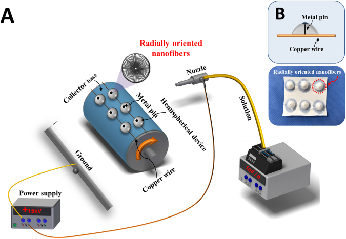
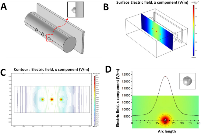
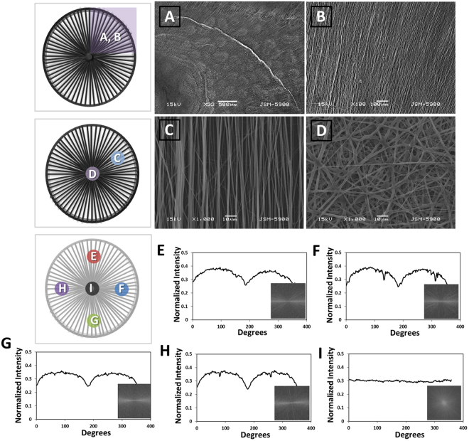
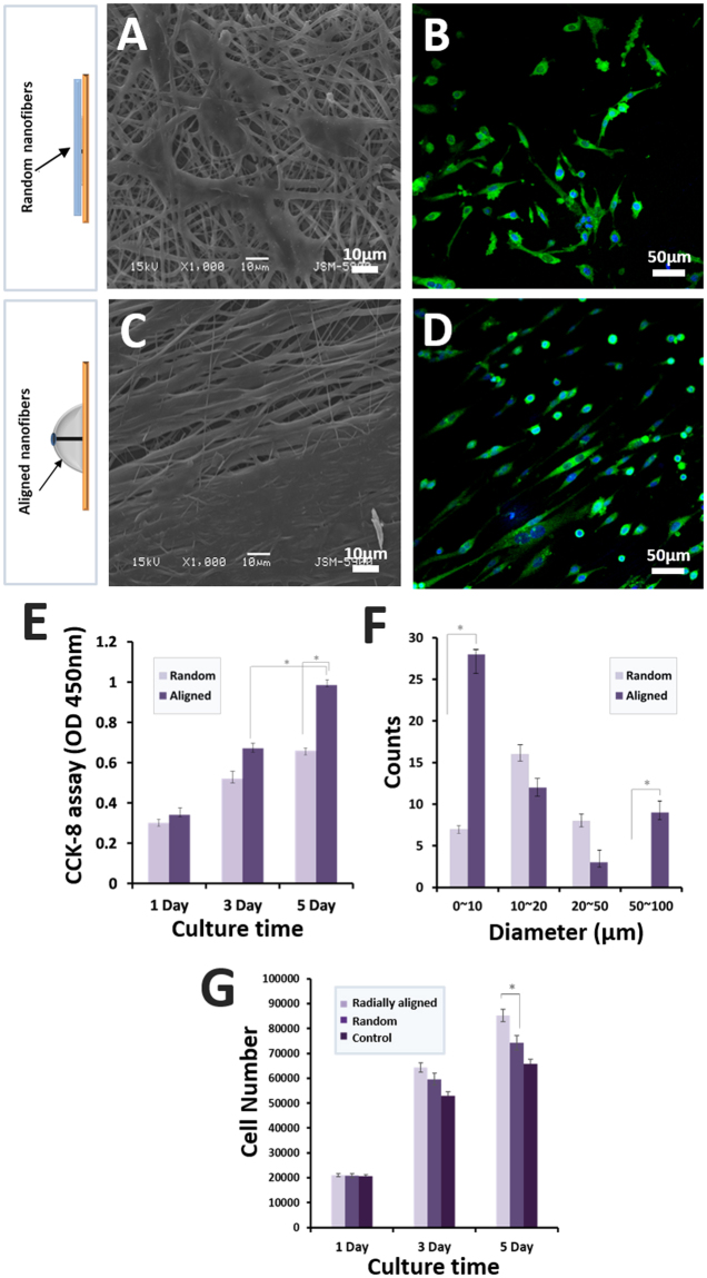
Tissue engineering has significantly contributed to the development of optimal treatments for individual injury sites based on their unique functional and histologic properties. Human organs and tissue have three-dimensional (3D) morphologies; for example, the morphology of the eye is a spherical shape. However, most conventional electrospinning equipment is only capable of fabricating a two-dimensional (2D) structured fibrous scaffold and no report is available on a 3D electrospinning method to fabricate a hemispherical scaffold to mimic the native properties of the cornea, including microscopic to macroscopic morphology and transparency. We proposed a novel electrospinning method using a single nonconductive hemispherical device and a metal pin. A designed peg-top shaped collector, a hemispherical nonconductive device with a metal pin in the center and copper wire forming a circle around at the edge was attached to a conventional conductive collector. A 3D hemispherical transparent scaffold with radially aligned nanofibers was successfully fabricated with the designed peg-top collector. In summary, our fabricated 3D electrospun scaffold is expected to be suitable for the treatment of injuries of ocular tissues owing to the hemispherical shape and radially aligned nanofibers which can guide the direction of the main collagen and cellular actin filament in the extracellular matrix.

摘要

组织工程学在根据个体损伤部位的独特功能和组织学特性,为开发最佳治疗方法方面做出了重大贡献。人类器官和组织具有三维(3D)形态;例如,眼睛的形态为球形。然而,大多数传统的静电纺丝设备仅能够制造二维(2D)结构的纤维支架,并且没有关于 3D 静电纺丝方法来制造半球形支架以模拟角膜固有特性的报告,包括微观到宏观形态和透明度。我们提出了一种使用单个非导电半球形装置和金属针的新型静电纺丝方法。一个设计的钉顶形状的收集器,一个带有中心金属针的半球形非导电装置,以及在边缘形成一个圆圈的铜丝,被连接到一个常规的导电收集器上。使用设计的钉顶收集器成功制造了具有径向排列纳米纤维的 3D 半球形透明支架。总之,由于半球形状和径向排列的纳米纤维,我们制造的 3D 静电纺丝支架有望适用于眼部组织损伤的治疗,因为这些纳米纤维可以引导细胞外基质中主要胶原蛋白和细胞肌动蛋白丝的方向。

https://cdn.ncbi.nlm.nih.gov/pmc/blobs/2213/5821851/dfc8b9481e83/41598_2018_21618_Fig8_HTML.jpg
https://cdn.ncbi.nlm.nih.gov/pmc/blobs/2213/5821851/3a6bad91ca01/41598_2018_21618_Fig1_HTML.jpg
https://cdn.ncbi.nlm.nih.gov/pmc/blobs/2213/5821851/0cfe69c82499/41598_2018_21618_Fig2_HTML.jpg
https://cdn.ncbi.nlm.nih.gov/pmc/blobs/2213/5821851/92ba4ebb4e4e/41598_2018_21618_Fig3_HTML.jpg
https://cdn.ncbi.nlm.nih.gov/pmc/blobs/2213/5821851/ee61c9775b1e/41598_2018_21618_Fig4_HTML.jpg
https://cdn.ncbi.nlm.nih.gov/pmc/blobs/2213/5821851/f33ff5497e3a/41598_2018_21618_Fig5_HTML.jpg
https://cdn.ncbi.nlm.nih.gov/pmc/blobs/2213/5821851/28401f74ca05/41598_2018_21618_Fig6_HTML.jpg
https://cdn.ncbi.nlm.nih.gov/pmc/blobs/2213/5821851/4eab6bf878c6/41598_2018_21618_Fig7_HTML.jpg
https://cdn.ncbi.nlm.nih.gov/pmc/blobs/2213/5821851/dfc8b9481e83/41598_2018_21618_Fig8_HTML.jpg

相似文献

1
Fabrication of transparent hemispherical 3D nanofibrous scaffolds with radially aligned patterns via a novel electrospinning method.采用新型静电纺丝方法制备具有径向排列图案的透明半球形 3D 纳米纤维支架。
Sci Rep. 2018 Feb 21;8(1):3424. doi: 10.1038/s41598-018-21618-0.
2
Fabrication of nanocomposite/nanofibrous functionally graded biomimetic scaffolds for osteochondral tissue regeneration.用于骨软骨组织再生的纳米复合/纳米纤维功能梯度仿生支架的制备。
J Biomed Mater Res A. 2021 Sep;109(9):1657-1669. doi: 10.1002/jbm.a.37161. Epub 2021 Mar 9.
3
Novel class of collector in electrospinning device for the fabrication of 3D nanofibrous structure for large defect load-bearing tissue engineering application.用于制造用于大缺陷承重组织工程应用的三维纳米纤维结构的静电纺丝装置中的新型收集器类别。
J Biomed Mater Res A. 2017 May;105(5):1535-1548. doi: 10.1002/jbm.a.35822. Epub 2017 Mar 21.
4
Mass production of nanofibrous extracellular matrix with controlled 3D morphology for large-scale soft tissue regeneration.大规模生产具有受控 3D 形态的纳米纤维细胞外基质,用于大规模软组织再生。
Tissue Eng Part C Methods. 2013 Jun;19(6):458-72. doi: 10.1089/ten.TEC.2012.0417. Epub 2012 Dec 12.
5
Design and fabrication of a nanofibrous polycaprolactone tubular nerve guide for peripheral nerve tissue engineering using a two-pole electrospinning system.使用两极静电纺丝系统设计和制造用于周围神经组织工程的纳米纤维聚己内酯管状神经导管。
Biomed Mater. 2016 Apr 12;11(2):025017. doi: 10.1088/1748-6041/11/2/025017.
6
Aligned conductive core-shell biomimetic scaffolds based on nanofiber yarns/hydrogel for enhanced 3D neurite outgrowth alignment and elongation.基于纳米纤维纱线/水凝胶的取向导电核壳仿生支架,用于增强 3D 神经突生长取向和延伸。
Acta Biomater. 2019 Sep 15;96:175-187. doi: 10.1016/j.actbio.2019.06.035. Epub 2019 Jun 29.
7
Three-Dimensional Hierarchical Nanofibrous Collagen Scaffold Fabricated Using Fibrillated Collagen and Pluronic F-127 for Regenerating Bone Tissue.采用原纤维化胶原蛋白和泊洛沙姆 F-127 制备用于骨组织再生的三维分层纳米纤维胶原支架
ACS Appl Mater Interfaces. 2018 Oct 24;10(42):35801-35811. doi: 10.1021/acsami.8b14088. Epub 2018 Oct 9.
8
Electrospun three-dimensional aligned nanofibrous scaffolds for tissue engineering.用于组织工程的静电纺丝三维定向纳米纤维支架。
Mater Sci Eng C Mater Biol Appl. 2018 Nov 1;92:995-1005. doi: 10.1016/j.msec.2018.06.065. Epub 2018 Jun 30.
9
Cell-matrix mechanical interaction in electrospun polymeric scaffolds for tissue engineering: Implications for scaffold design and performance.用于组织工程的电纺聚合物支架中的细胞-基质机械相互作用:对支架设计和性能的影响。
Acta Biomater. 2017 Mar 1;50:41-55. doi: 10.1016/j.actbio.2016.12.034. Epub 2016 Dec 21.
10
Three-dimensional electrospun nanofibrous scaffolds for bone tissue engineering.用于骨组织工程的三维电纺纳米纤维支架
J Biomed Mater Res B Appl Biomater. 2020 May;108(4):1311-1321. doi: 10.1002/jbm.b.34479. Epub 2019 Aug 22.

引用本文的文献

1
BMSC-NFMC Model for Vascular Regulation and Interface Integration in Osteochondral Regeneration.用于骨软骨再生中血管调节和界面整合的骨髓间充质干细胞-纳米纤维微载体复合物模型
Adv Sci (Weinh). 2025 Sep;12(33):e05222. doi: 10.1002/advs.202505222. Epub 2025 Jun 23.
2
3D Electrospun Synthetic Extracellular Matrix for Tissue Regeneration.用于组织再生的3D电纺合成细胞外基质
Small Sci. 2021 May 25;1(7):2100003. doi: 10.1002/smsc.202100003. eCollection 2021 Jul.
3
Fabrication of 3D Polycaprolactone Macrostructures by 3D Electrospinning.3D 静电纺丝法制备 3D 聚己内酯宏观结构

本文引用的文献

1
Electrospun anisotropic architectures and porous structures for tissue engineering.用于组织工程的电纺各向异性结构和多孔结构
J Mater Chem B. 2015 Jul 21;3(27):5389-5410. doi: 10.1039/c5tb00472a. Epub 2015 Jun 10.
2
Electrospun Scaffolds for Corneal Tissue Engineering: A Review.用于角膜组织工程的电纺支架:综述
Materials (Basel). 2016 Jul 27;9(8):614. doi: 10.3390/ma9080614.
3
Highly defined 3D printed chitosan scaffolds featuring improved cell growth.具有改进细胞生长性能的高分辨率 3D 打印壳聚糖支架。
ACS Biomater Sci Eng. 2024 Aug 12;10(8):5336-5351. doi: 10.1021/acsbiomaterials.4c00302. Epub 2024 May 22.
4
Investigating the effect of poly (ɛ-caprolactone) nanofibers scaffolds with random, unidirectionally, and radially aligned morphologies on the Fibroblast cell's attachment and growth behavior.研究具有随机、单向和径向排列形态的聚(ε-己内酯)纳米纤维支架对成纤维细胞附着和生长行为的影响。
Turk J Chem. 2022 Nov 8;47(1):54-62. doi: 10.55730/1300-0527.3516. eCollection 2023.
5
Cell-based Therapies for Corneal and Retinal Disorders.基于细胞的角膜和视网膜疾病疗法。
Stem Cell Rev Rep. 2023 Nov;19(8):2650-2682. doi: 10.1007/s12015-023-10623-0. Epub 2023 Sep 14.
6
Three-Dimensional Scaffolds for Bone Tissue Engineering.用于骨组织工程的三维支架
Bioengineering (Basel). 2023 Jun 25;10(7):759. doi: 10.3390/bioengineering10070759.
7
Detection of Limbal Stem Cells Adhered to Melt Electrospun Silk Fibroin and Gelatin-Modified Polylactic Acid Scaffolds.检测附着于熔体静电纺丝丝素蛋白和明胶改性聚乳酸支架上的角膜缘干细胞。
Polymers (Basel). 2023 Feb 3;15(3):777. doi: 10.3390/polym15030777.
8
Periodontal Ligament-Mimetic Fibrous Scaffolds Regulate YAP-Associated Fibroblast Behaviors and Promote Regeneration of Periodontal Defect in Relation to the Scaffold Topography.牙周韧带仿生纤维支架通过调节 YAP 相关成纤维细胞行为并与支架形貌相关促进牙周缺损再生。
ACS Appl Mater Interfaces. 2023 Jan 11;15(1):599-616. doi: 10.1021/acsami.2c18893. Epub 2022 Dec 28.
9
Electrospun hybrid nanofibers: Fabrication, characterization, and biomedical applications.静电纺丝复合纳米纤维:制备、表征及生物医学应用。
Front Bioeng Biotechnol. 2022 Dec 1;10:986975. doi: 10.3389/fbioe.2022.986975. eCollection 2022.
10
Bioprinted Membranes for Corneal Tissue Engineering: A Review.用于角膜组织工程的生物打印膜:综述
Pharmaceutics. 2022 Dec 14;14(12):2797. doi: 10.3390/pharmaceutics14122797.
Biomed Mater. 2017 Jul 12;12(4):045009. doi: 10.1088/1748-605X/aa7692.
4
Collagen density gradient on three-dimensional printed poly(ε-caprolactone) scaffolds for interface tissue engineering.三维打印聚己内酯支架上的胶原密度梯度用于界面组织工程。
J Tissue Eng Regen Med. 2018 Feb;12(2):321-329. doi: 10.1002/term.2457. Epub 2017 Aug 17.
5
Tissue-engineered cornea constructed with compressed collagen and laser-perforated electrospun mat.采用压缩胶原和激光穿孔的静电纺丝垫构建组织工程角膜。
Sci Rep. 2017 Apr 20;7(1):970. doi: 10.1038/s41598-017-01072-0.
6
Controlling the 3D architecture of Self-Lifting Auto-generated Tissue Equivalents (SLATEs) for optimized corneal graft composition and stability.控制自提升自动生成组织等效物(SLATEs)的3D结构,以优化角膜移植的组成和稳定性。
Biomaterials. 2017 Mar;121:205-219. doi: 10.1016/j.biomaterials.2016.12.023. Epub 2016 Dec 23.
7
Adhesion and metabolic activity of human corneal cells on PCL based nanofiber matrices.人角膜细胞在聚己内酯基纳米纤维基质上的黏附及代谢活性
Mater Sci Eng C Mater Biol Appl. 2017 Feb 1;71:764-770. doi: 10.1016/j.msec.2016.10.058. Epub 2016 Oct 26.
8
3D printed alendronate-releasing poly(caprolactone) porous scaffolds enhance osteogenic differentiation and bone formation in rat tibial defects.3D打印的释放阿仑膦酸盐的聚己内酯多孔支架增强大鼠胫骨缺损处的成骨分化和骨形成。
Biomed Mater. 2016 Sep 29;11(5):055005. doi: 10.1088/1748-6041/11/5/055005.
9
Nature-Derived Aloe Vera Gel Blended Silk Fibroin Film Scaffolds for Cornea Endothelial Cell Regeneration and Transplantation.天然芦荟凝胶与丝素蛋白复合膜支架用于角膜内皮细胞再生和移植。
ACS Appl Mater Interfaces. 2016 Jun 22;8(24):15160-8. doi: 10.1021/acsami.6b04901. Epub 2016 Jun 8.
10
Formation of Highly Aligned Collagen Nanofibers by Continuous Cyclic Stretch of a Collagen Hydrogel Sheet.通过胶原水凝胶片的连续循环拉伸形成高度排列的胶原纳米纤维。
Macromol Biosci. 2016 Jul;16(7):995-1000. doi: 10.1002/mabi.201600068. Epub 2016 May 2.