Department of Neuroscience, Biomedicine and Movement, Human Anatomy and Histology Section, University of Verona, Verona I-37134, Italy.
Department of Medicine, Gastroenterology Section, University of Verona, Verona I-37134, Italy.
World J Gastroenterol. 2018 Feb 21;24(7):775-793. doi: 10.3748/wjg.v24.i7.775.
To investigate by immunostaining glucose transporter expression in human colorectal mucosa in controls and patients with inflammatory bowel disease (IBD).
Colorectal samples were obtained from patients undergoing lower endoscopic colonoscopy or recto-sigmoidoscopy. Patients diagnosed with ulcerative colitis ( = 18) or Crohn's disease ( = 10) and scheduled for diagnostic colonoscopy were enrolled. Patients who underwent colonoscopy for prevention screening of colorectal cancer or were followed-up after polypectomy or had a history of lower gastrointestinal symptoms were designated as the control group (CTRL, = 16). Inflammatory status of the mucosa at the sampling site was evaluated histologically and/or endoscopically. A total of 147 biopsies of colorectal mucosa were collected and processed for immunohistochemistry analysis. The expression of GLUT2, SGLT1, and GLUT5 glucose transporters was investigated using immunoperoxidase labeling. To compare immunoreactivity of GLUT5 and LYVE-1, which is a marker for lymphatic vessel endothelium, double-labeled confocal microscopy was used.
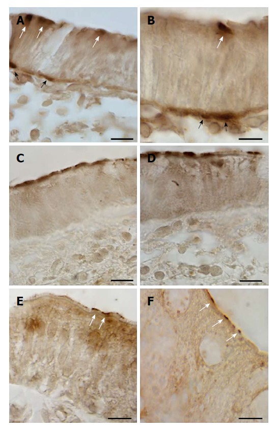
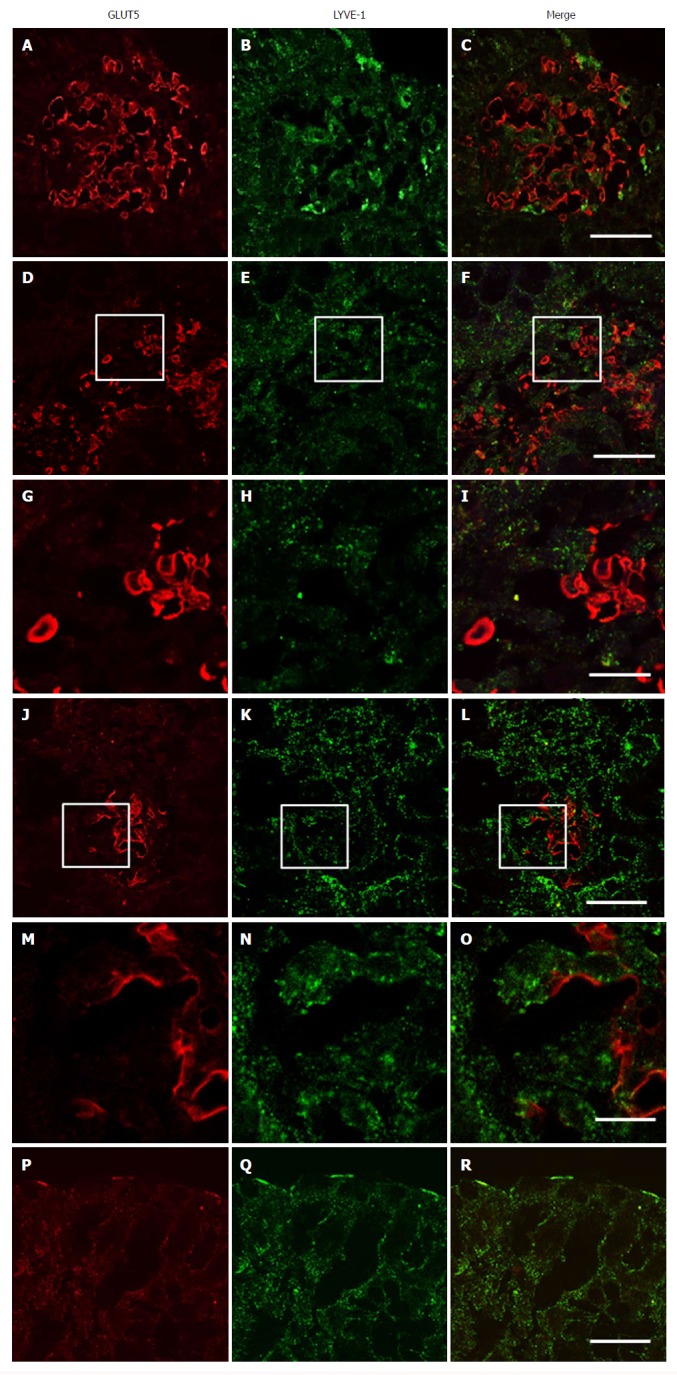
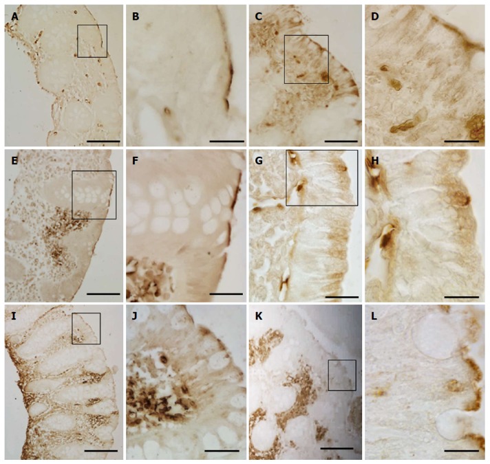
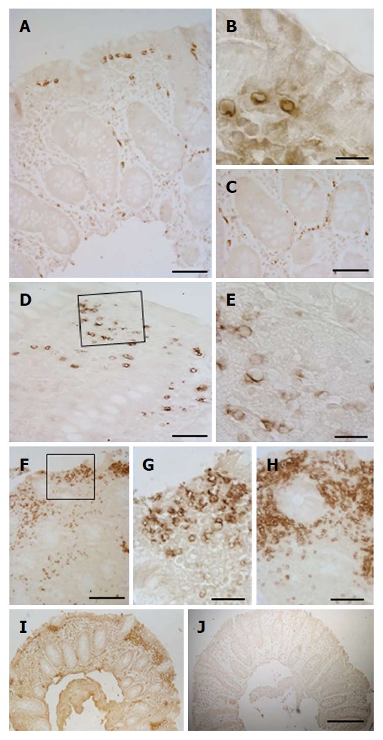
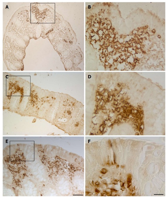
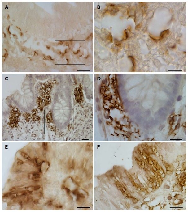
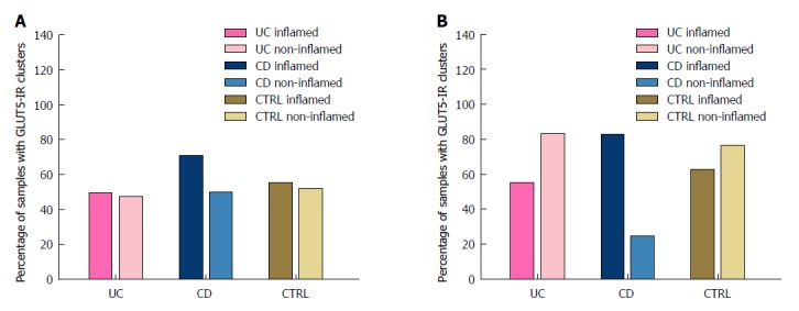
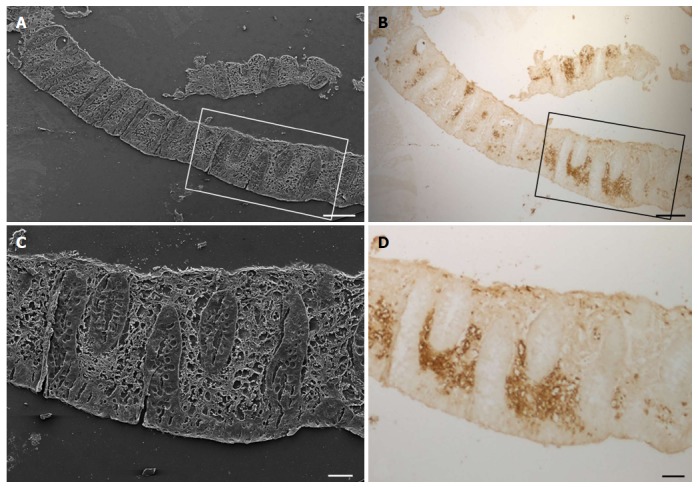
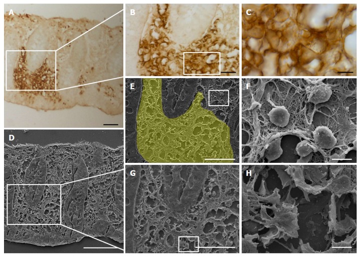
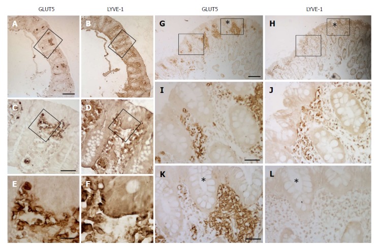
Immunohistochemical analysis revealed that GLUT2, SGLT1, and GLUT5 were expressed only in short epithelial portions of the large intestinal mucosa. No important differences were observed in glucose transporter expression between the samples obtained from the different portions of the colorectal tract and between the different patient groups. Unexpectedly, GLUT5 expression was also identified in vessels, mainly concentrated in specific areas where the vessels were clustered. Immunostaining with LYVE-1 and GLUT5 antibodies revealed that GLUT5-immunoreactive (-IR) clusters of vessels were concentrated in areas internal to those that were LYVE-1 positive. GLUT5 and LYVE-1 did not appear to be colocalized but rather showed a close topographical relationship on the endothelium. Based on their LYVE-1 expression, GLUT5-IR vessels were identified as lymphatic. Both inflamed and non-inflamed mucosal colorectal tissue biopsies from the IBD and CTRL patients showed GLUT5-IR clusters of lymphatic vessels.
Glucose transporter immunoreactivity is present in colorectal mucosa in controls and IBD patients. GLUT5 expression is also associated with lymphatic vessels. This novel finding aids in the characterization of lymphatic vasculature in IBD patients.
通过免疫组化染色研究葡萄糖转运体在人类结直肠黏膜中的表达,以观察其在对照和炎症性肠病(IBD)患者中的变化。
从接受下内窥镜结肠镜或直肠乙状结肠镜检查的患者中获得结直肠样本。招募诊断为溃疡性结肠炎(= 18)或克罗恩病(= 10)并计划接受诊断性结肠镜检查的患者。将因预防结直肠癌筛查、息肉切除后随访或有下消化道症状史而接受结肠镜检查的患者指定为对照组(CTRL,= 16)。通过组织学和/或内窥镜检查评估取样部位的黏膜炎症状态。共收集并处理了 147 例结直肠黏膜活检标本,用于免疫组织化学分析。使用免疫过氧化物酶标记法研究 GLUT2、SGLT1 和 GLUT5 葡萄糖转运体的表达。为了比较 GLUT5 和 LYVE-1(淋巴管内皮标志物)的免疫反应性,使用双标记共聚焦显微镜。
免疫组织化学分析显示,GLUT2、SGLT1 和 GLUT5 仅在大肠黏膜的短上皮部分表达。不同部位和不同患者组之间,葡萄糖转运体的表达没有明显差异。出乎意料的是,GLUT5 的表达也存在于血管中,主要集中在血管聚集的特定区域。LYVE-1 和 GLUT5 抗体的免疫染色显示,GLUT5 免疫反应性(-IR)血管簇集中在 LYVE-1 阳性区域内部。GLUT5 和 LYVE-1 似乎没有共定位,而是在内皮上表现出密切的拓扑关系。根据 LYVE-1 的表达,GLUT5-IR 血管被鉴定为淋巴管。来自 IBD 和 CTRL 患者的炎症和非炎症性结直肠组织活检均显示 GLUT5-IR 淋巴管簇。
葡萄糖转运体在对照和 IBD 患者的结直肠黏膜中均有表达。GLUT5 的表达也与淋巴管有关。这一新发现有助于 IBD 患者中淋巴管血管的特征描述。