Molecular Imaging Program at Stanford (MIPS), Department of Radiology, Stanford University, Stanford, CA, 94305, USA.
Department of Neurology and Neurological Sciences, Stanford University, Stanford, CA, 94305, USA.
J Neuroinflammation. 2018 Feb 22;15(1):55. doi: 10.1186/s12974-018-1080-1.
The cystine/glutamate antiporter (xc-) has been implicated in several neurological disorders and, specifically, in multiple sclerosis (MS) as a mediator of glutamate excitotoxicity and proinflammatory immune responses. We aimed to evaluate an xc-specific positron emission tomography (PET) radiotracer, (4S)-4-(3-[F]fluoropropyl)-L-glutamate ([F]FSPG), for its ability to allow non-invasive monitoring of xc- activity in a mouse model of MS.
Experimental autoimmune encephalomyelitis (EAE) was induced in C57BL/6 mice by subcutaneous injection of myelin oligodendrocyte glycoprotein (MOG) peptide in complete Freund's adjuvant (CFA) followed by pertussis toxin. Control mice received CFA emulsion and pertussis toxin without MOG peptide, while a separate cohort of naïve mice received no treatment. PET studies were performed to investigate the kinetics and distribution of [F]FSPG in naïve, control, pre-symptomatic, and symptomatic EAE mice, compared to F-fluorodeoxyglucose ([F]FDG). After final PET scans, each mouse was perfused and radioactivity in dissected tissues was measured using a gamma counter. Central nervous system (CNS) tissues were further analyzed using ex vivo autoradiography or western blot. [F]FSPG uptake in human monocytes, and T cells pre- and post-activation was investigated in vitro.
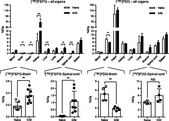
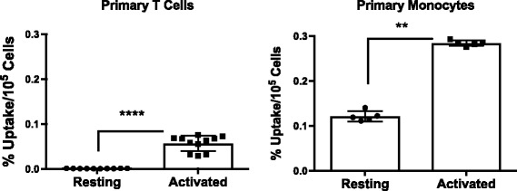
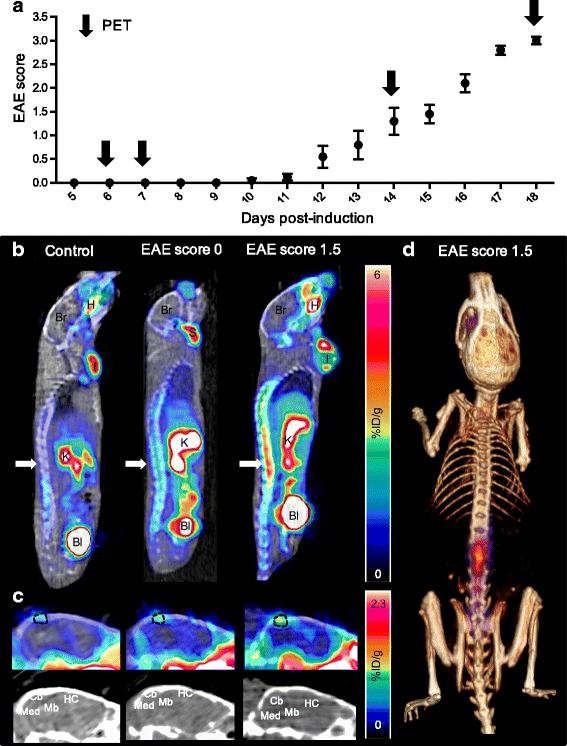
[F]FSPG was found to be more sensitive than [F]FDG at detecting pathological changes in the spinal cord and brain of EAE mice. Even before clinical signs of disease, a small but significant increase in [F]FSPG signal was observed in the spinal cord of EAE mice compared to controls. This increase in PET signal became more pronounced in symptomatic EAE mice and was confirmed by ex vivo biodistribution and autoradiography. Likewise, in the brain of symptomatic EAE mice, [F]FSPG uptake was significantly higher than controls, with the largest changes observed in the cerebellum. Western blot analyses of CNS tissues revealed a significant correlation between light chain of xc- (xCT) protein levels, the subunit of xc- credited with its transporter activity, and [F]FSPG-PET signal. In vitro [F]FSPG uptake studies suggest that both activated monocytes and T cells contribute to the observed in vivo PET signal.
These data highlight the promise of [F]FSPG-PET as a technique to provide insights into neuroimmune interactions in MS and the in vivo role of xc- in the development and progression of this disease, thus warranting further investigation.
胱氨酸/谷氨酸反向转运体 (xc-) 与多种神经系统疾病有关,特别是多发性硬化症 (MS),作为谷氨酸兴奋性毒性和促炎免疫反应的介质。我们旨在评估一种 xc-特异性正电子发射断层扫描 (PET) 示踪剂,(4S)-4-(3-[F] 氟丙基)-L-谷氨酸 ([F]FSPG),用于在 MS 的小鼠模型中允许非侵入性监测 xc- 活性。
通过在完全弗氏佐剂 (CFA) 中皮下注射髓鞘少突胶质细胞糖蛋白 (MOG) 肽,然后给予百日咳毒素,在 C57BL/6 小鼠中诱导实验性自身免疫性脑脊髓炎 (EAE)。对照小鼠接受 CFA 乳液和百日咳毒素而没有 MOG 肽,而另一组未处理的对照小鼠接受无治疗。进行 PET 研究以研究 [F]FSPG 在未处理、对照、前驱和症状性 EAE 小鼠中的动力学和分布,与 F-氟脱氧葡萄糖 ([F]FDG) 进行比较。在最后一次 PET 扫描后,使用伽马计数器测量每个小鼠的放射性。使用放射性自显影或 Western blot 进一步分析中枢神经系统 (CNS) 组织。在体外研究了 [F]FSPG 在单核细胞和 T 细胞激活前后的摄取。
与 [F]FDG 相比,[F]FSPG 更能检测 EAE 小鼠脊髓和大脑的病理变化。甚至在疾病的临床症状出现之前,与对照相比,EAE 小鼠的脊髓中就观察到 [F]FSPG 信号的小但显著增加。在症状性 EAE 小鼠中,这种 PET 信号的增加更加明显,并通过离体生物分布和放射性自显影得到证实。同样,在症状性 EAE 小鼠的大脑中,[F]FSPG 的摄取明显高于对照,小脑的变化最大。CNS 组织的 Western blot 分析显示,xc-的轻链 (xCT) 蛋白水平与 xc-的亚基之间存在显著相关性,xc-被认为具有其转运体活性。体外 [F]FSPG 摄取研究表明,激活的单核细胞和 T 细胞都有助于观察到的体内 PET 信号。
这些数据强调了 [F]FSPG-PET 作为一种提供多发性硬化症中神经免疫相互作用以及 xc- 在该疾病发展和进展中的体内作用的技术的潜力,因此值得进一步研究。