The Russell H. Morgan Department of Radiology and Radiological Science, Johns Hopkins Medical Institutions, Baltimore, MD, USA.
, JHOC 3140E, 601 N. Caroline Street, Baltimore, MD, 21287, USA.
Abdom Radiol (NY). 2018 Feb;43(2):445-456. doi: 10.1007/s00261-017-1338-6.
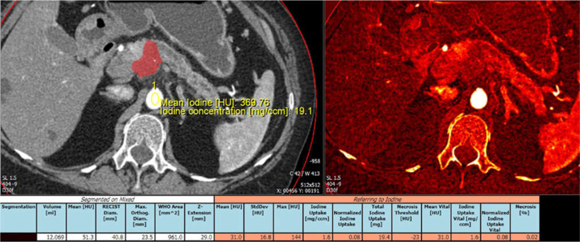
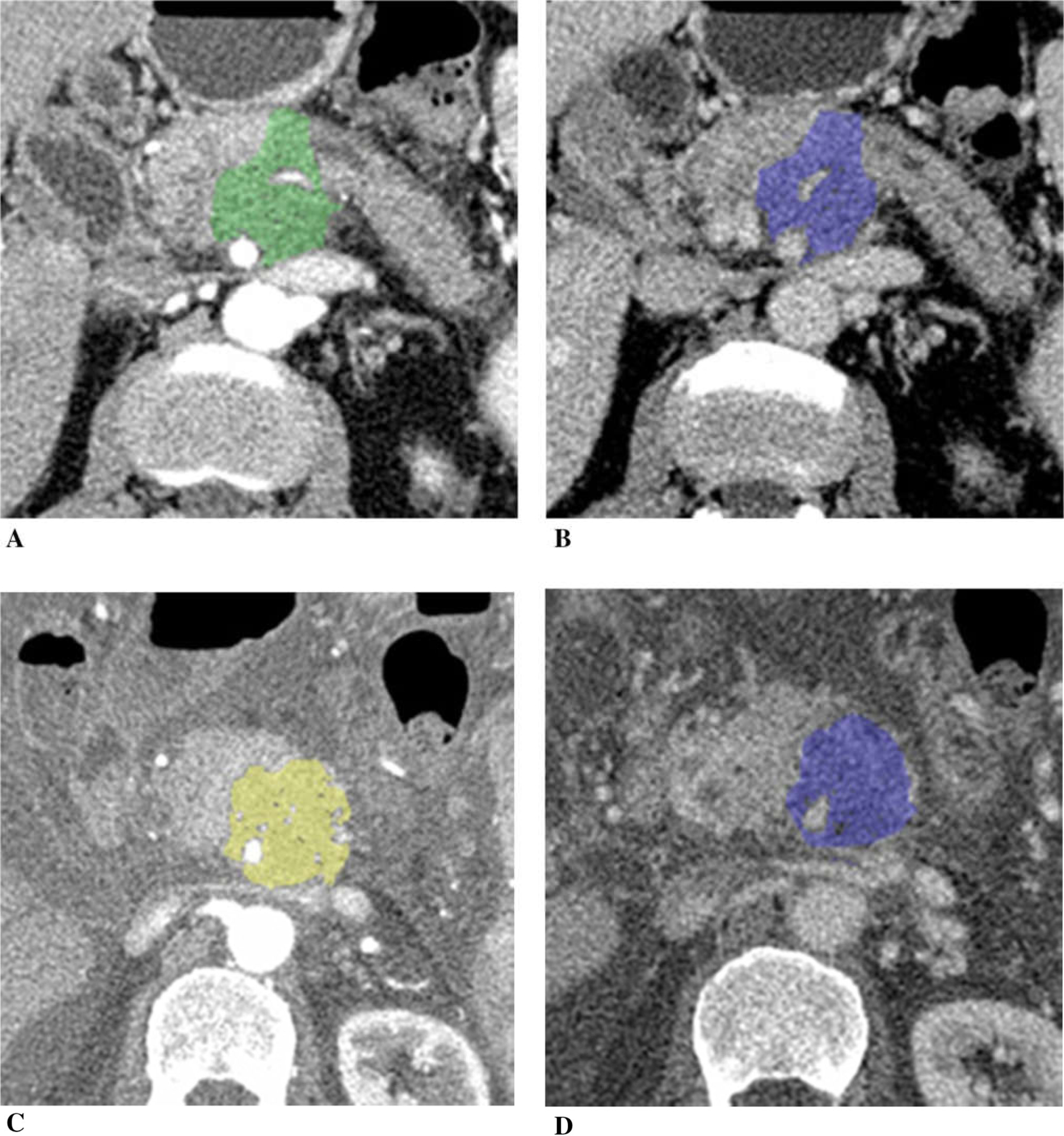
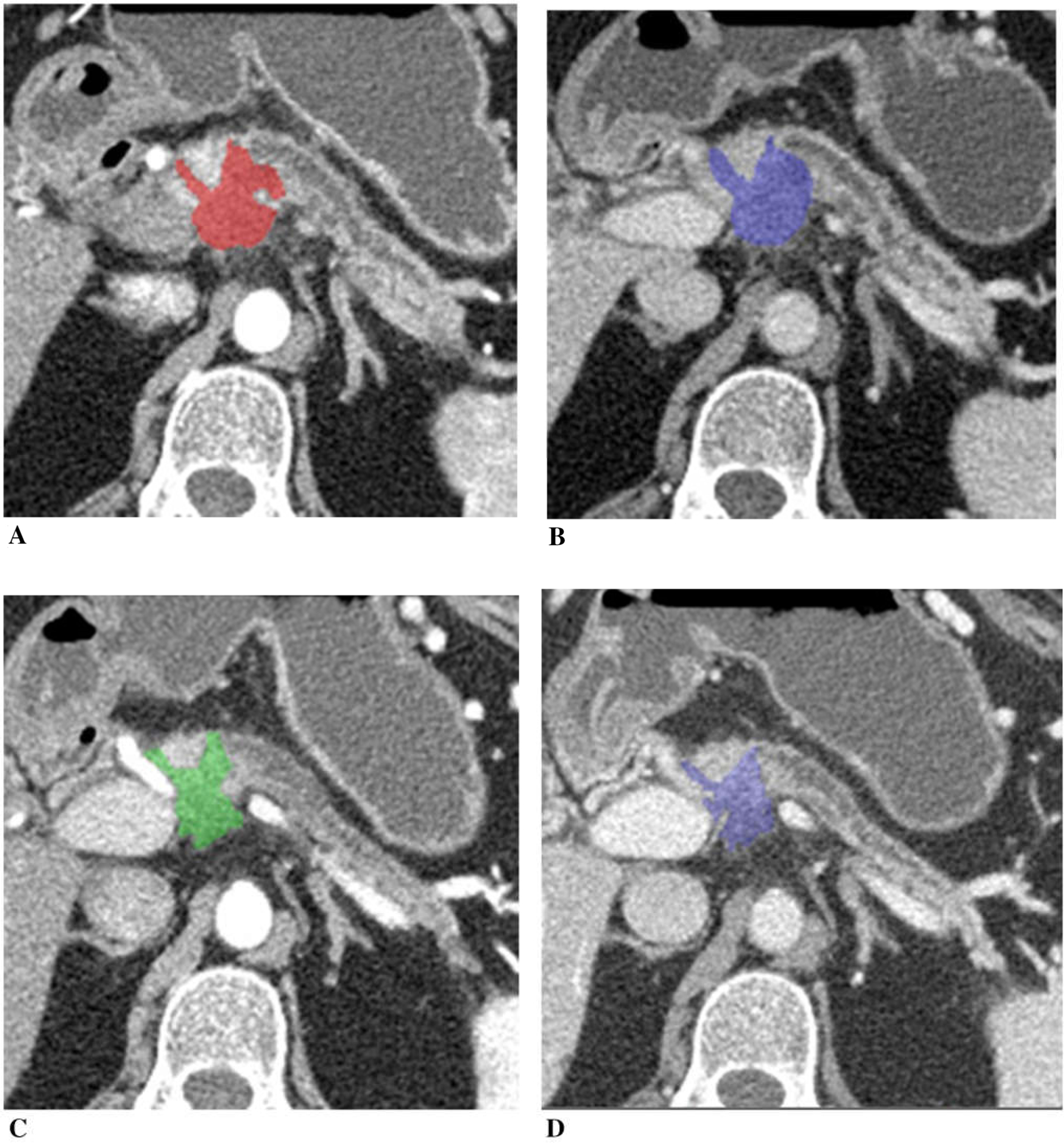
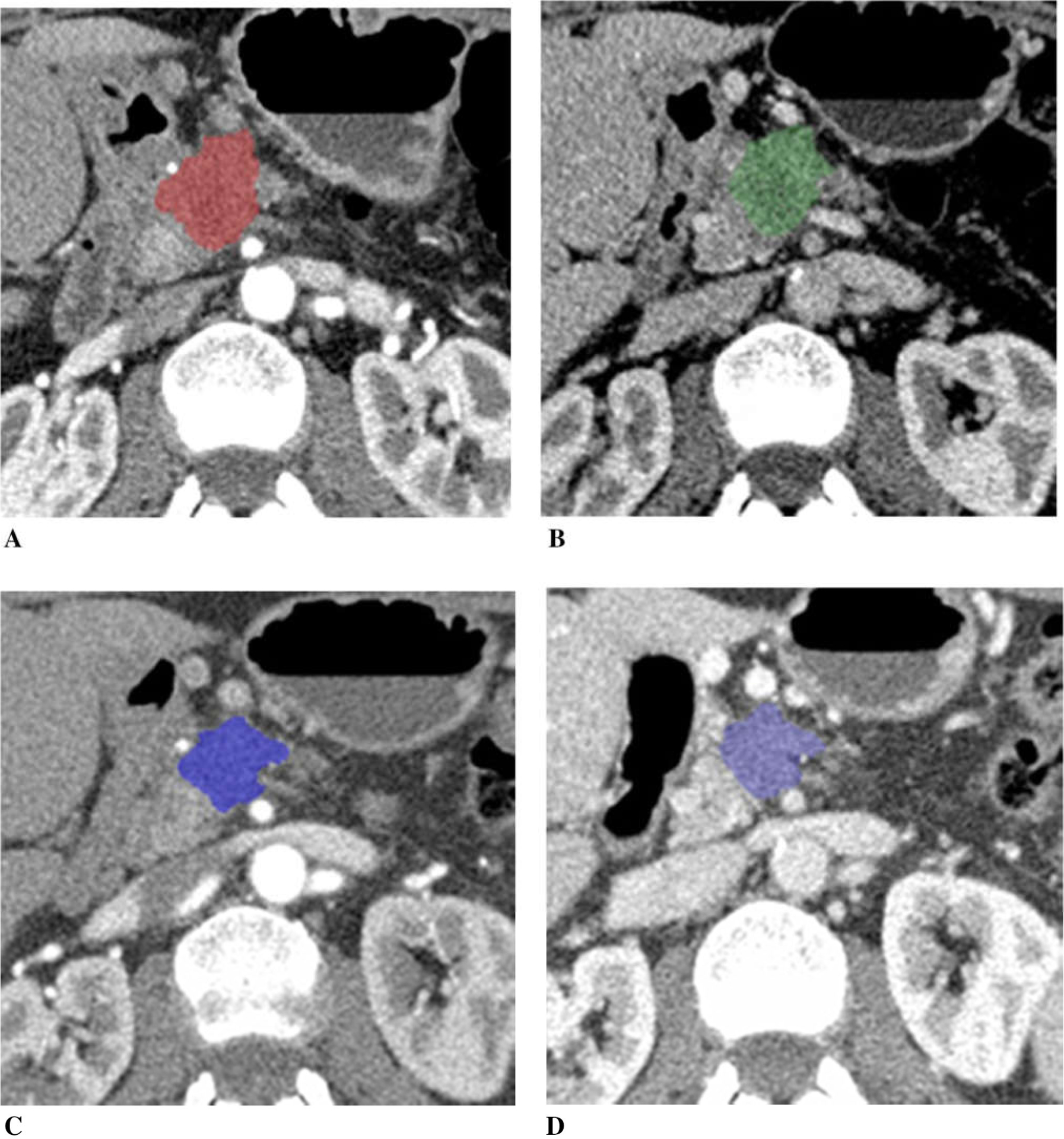
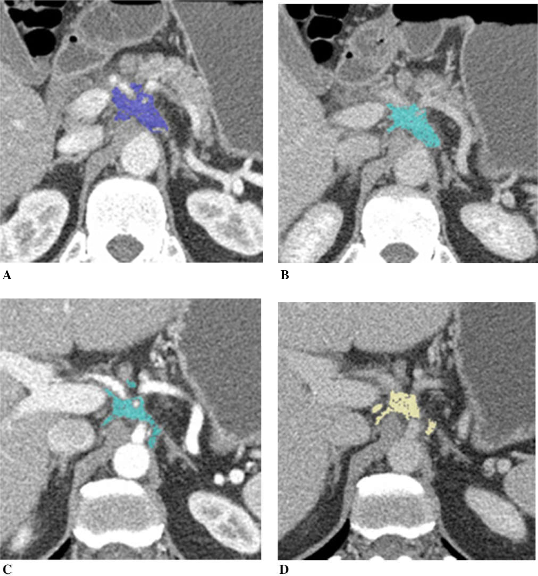
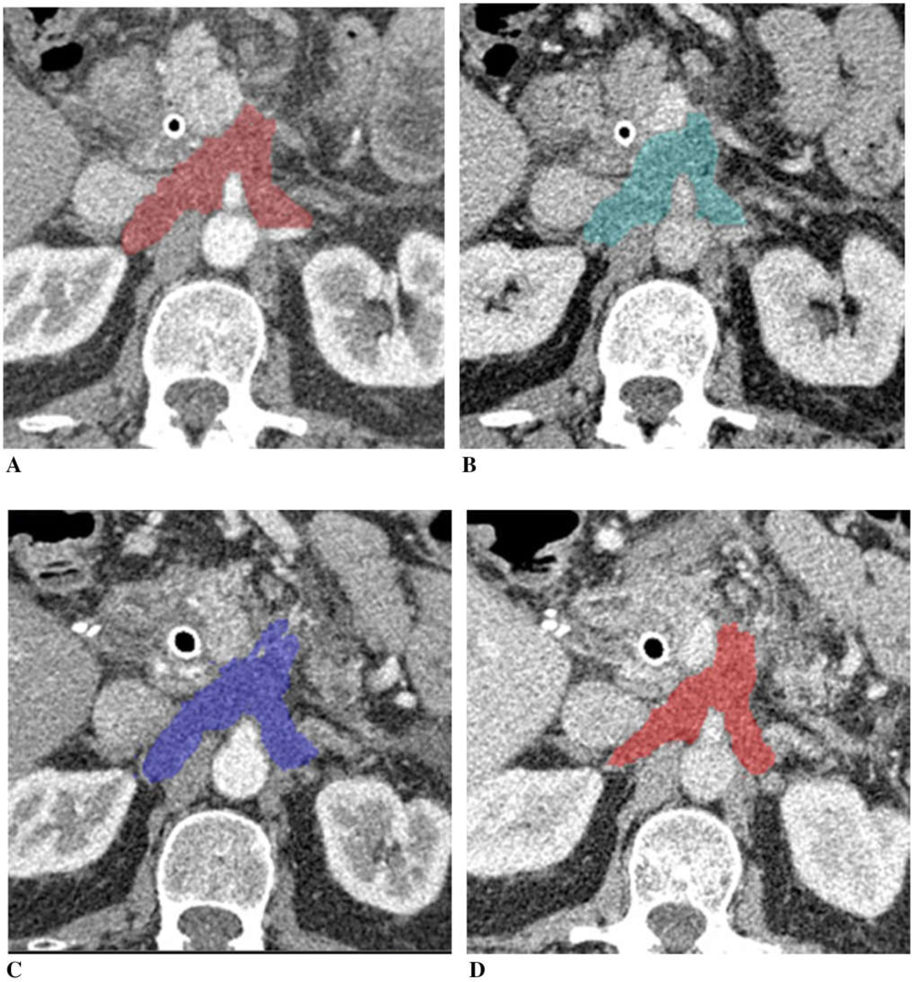
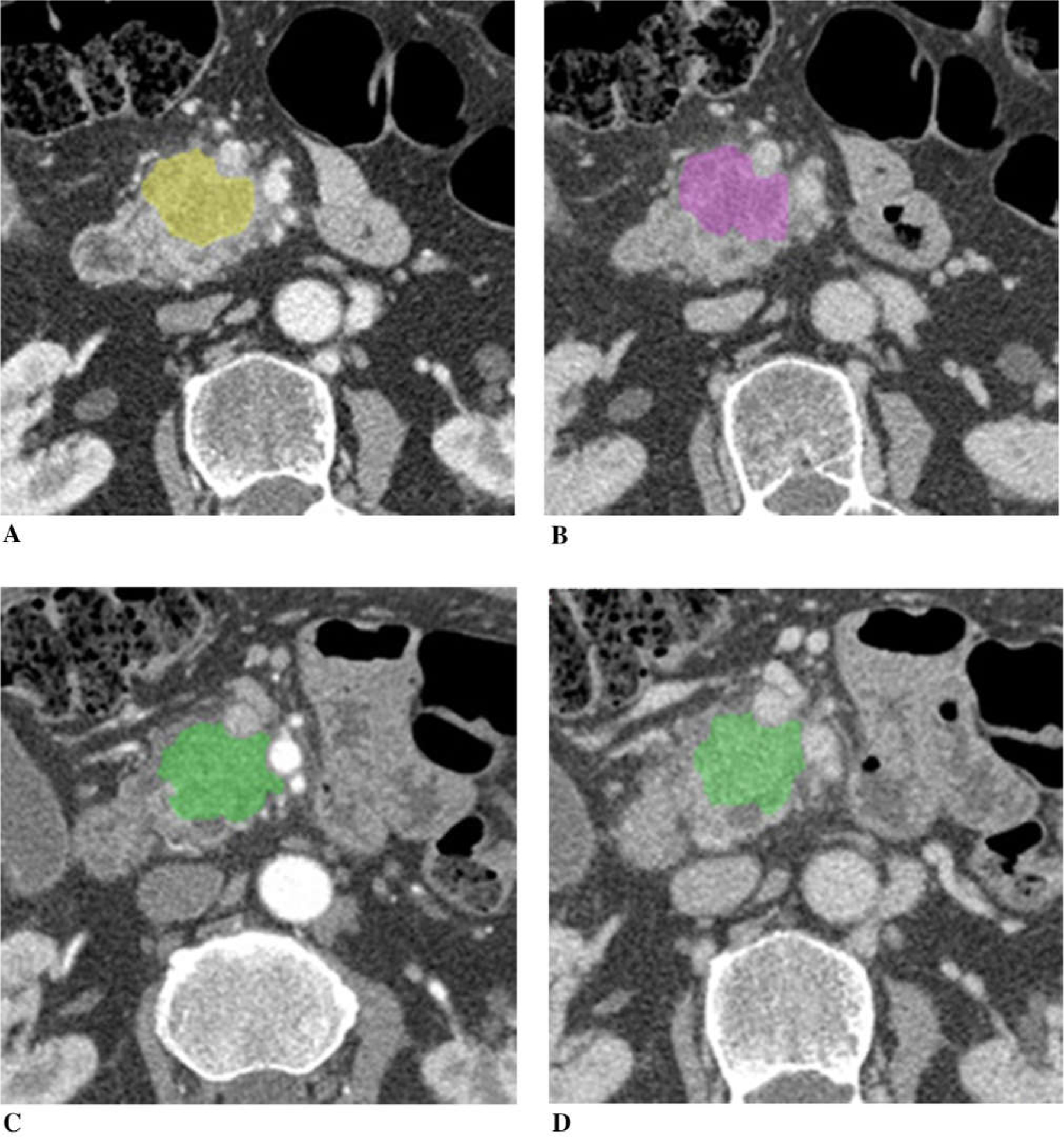
Pancreatic cancer remains a major health problem, and only less than 20% of patients have resectable disease at the time of initial diagnosis. Systemic chemotherapy is often used in the patients with borderline resectable, locally advanced unresectable disease and metastatic disease. CT is often used to assess for therapeutic response; however, conventional imaging including CT may not correctly reflect treatment response after chemotherapy. Dual-energy (DE) CT can acquire datasets at two different photon spectra in a single CT acquisition, and permits separating materials and extract iodine by applying a material decomposition algorithm. Quantitative iodine mapping may have an added value over conventional CT imaging for monitoring the treatment effects in patients with pancreatic cancer and potentially serve as a unique biomarker for treatment response. In this pictorial essay, we will review the technique for iodine quantification of pancreatic cancer by DECT and discuss our observations of iodine quantification at baseline and after systemic chemotherapy with conventional cytotoxic agents, and illustrate example cases.
胰腺癌仍然是一个主要的健康问题,只有不到 20%的患者在初始诊断时患有可切除的疾病。对于边界可切除、局部晚期不可切除疾病和转移性疾病的患者,常采用全身化疗。CT 常用于评估治疗反应;然而,包括 CT 在内的常规影像学检查可能无法正确反映化疗后的治疗反应。双能(DE)CT 可以在单次 CT 采集过程中获取两个不同光子能谱的数据集,并通过应用物质分解算法来分离物质和提取碘。与常规 CT 成像相比,定量碘图可能对监测胰腺癌患者的治疗效果具有附加价值,并可能成为治疗反应的独特生物标志物。在本影像学文章中,我们将回顾 DECT 对胰腺癌碘定量的技术,并讨论我们对基线和全身化疗后常规细胞毒性药物碘定量的观察结果,并举例说明。