Keppel Marc, Davoudi Eva, Gätgens Cornelia, Frunzke Julia
Institute of Bio- and Geosciences, IBG-1: Biotechnology, Forschungszentrum Jülich, Jülich, Germany.
Front Microbiol. 2018 Feb 9;9:183. doi: 10.3389/fmicb.2018.00183. eCollection 2018.
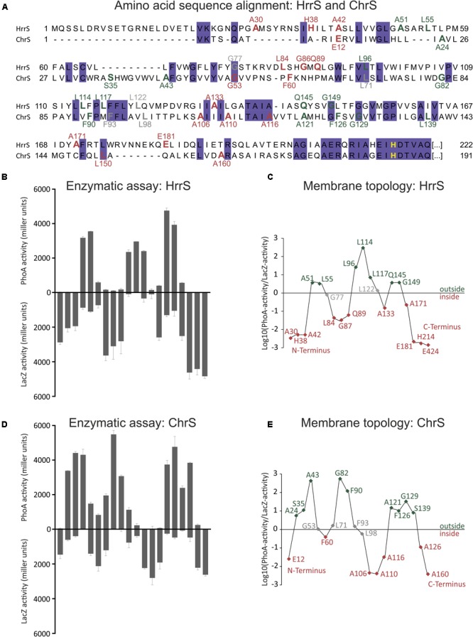
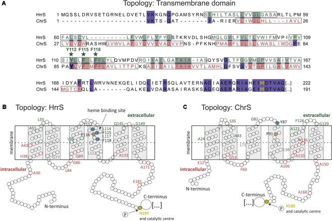
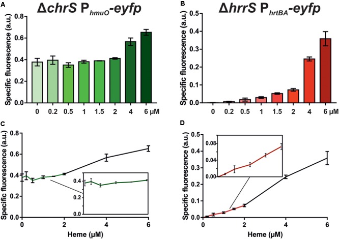
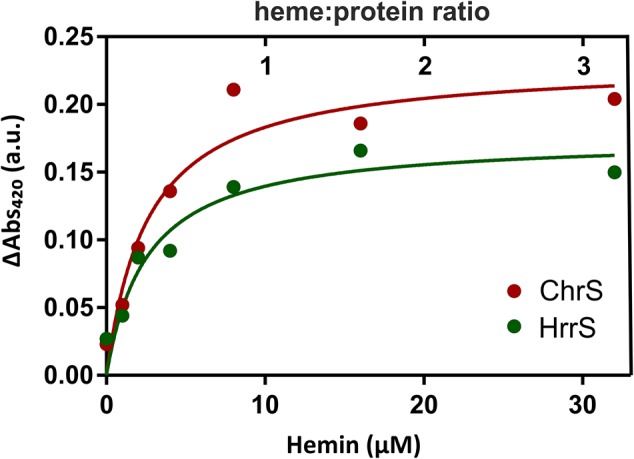
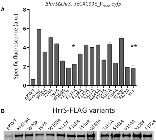
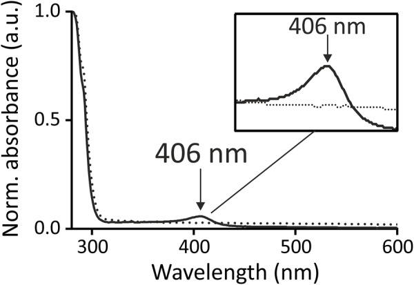
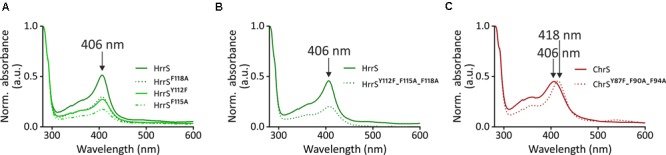
The HrrSA and the ChrSA two-component systems play a central role in the coordination of heme homeostasis in the Gram-positive soil bacterium and the prominent pathogen , both members of the . In this study, we have performed a comparative analysis of the membrane topology and heme-binding characteristics of the histidine kinases HrrS and ChrS of . While the cytoplasmic catalytic domains are highly conserved between HrrS and ChrS, the N-terminal sensing parts share only minor sequence similarity. PhoA and LacZ fusions of the N-terminal sensor domains of HrrS and ChrS revealed that both proteins are embedded into the cytoplasmic membrane via six α-helices. Although the overall membrane topology appeared to be conserved, target gene profiling indicated a higher sensitivity of the ChrS system to low heme levels (< 1 μM). , solubilized and purified full-length proteins bound heme in a 1:1 stoichiometry per monomer. Alanine-scanning of conserved amino acid residues in the N-terminal sensor domain revealed three aromatic residues (Y, F, and F), which apparently contribute to heme binding of HrrS. Exchange of either one or all three residues resulted in an almost abolished heme binding of HrrS . In contrast, ChrS mutants only displayed a red shift of the soret band from 406 to 418 nm suggesting an altered set of ligands in the triple mutant. In line with target gene profiling, these studies suggest distinct differences in the heme-protein interface of HrrS and ChrS. Since the membrane topology mapping displayed no extensive loop regions and alanine-scanning revealed potential heme-binding residues in α-helix number four, we propose an intramembrane sensing mechanism for both proteins. Overall, we present a first comparative analysis of the ChrS and HrrS kinases functioning as transient heme sensors in the .
HrrSA和ChrSA双组分系统在革兰氏阳性土壤细菌以及该菌纲中的重要病原体的血红素稳态协调中起着核心作用。在本研究中,我们对[细菌名称]的组氨酸激酶HrrS和ChrS的膜拓扑结构和血红素结合特性进行了比较分析。虽然HrrS和ChrS的细胞质催化结构域高度保守,但N端传感部分仅具有较小的序列相似性。HrrS和ChrS的N端传感器结构域的PhoA和LacZ融合表明,这两种蛋白质都通过六个α螺旋嵌入细胞质膜。尽管整体膜拓扑结构似乎是保守的,但靶基因分析表明ChrS系统对低血红素水平(<1μM)具有更高的敏感性。[具体实验步骤或结果],溶解并纯化的全长蛋白质以每单体1:1的化学计量比结合血红素。对N端传感器结构域中保守氨基酸残基进行丙氨酸扫描,发现三个芳香族残基(Y、F和F),它们显然有助于HrrS的血红素结合。单个或所有三个残基的交换导致HrrS的血红素结合几乎完全丧失。相比之下,ChrS突变体仅显示索雷特带从406nm红移至418nm,表明三重突变体中配体组发生了改变。与靶基因分析一致,这些研究表明HrrS和ChrS的血红素-蛋白质界面存在明显差异。由于膜拓扑图谱未显示广泛的环区,且丙氨酸扫描揭示了α螺旋四号中的潜在血红素结合残基,我们提出这两种蛋白质均存在膜内传感机制。总体而言,我们首次对在[细菌名称]中作为瞬时血红素传感器发挥作用的ChrS和HrrS激酶进行了比较分析。