Key Laboratory of Transplant Engineering and Immunology, National Health and Family Planning Commission (NHFPC), West China Hospital, Sichuan University, Chengdu, Sichuan 610041, P.R. China.
Department of Nephrology, West China Hospital, Sichuan University, Chengdu, Sichuan 610041, P.R. China.
Int J Mol Med. 2018 May;41(5):2629-2639. doi: 10.3892/ijmm.2018.3501. Epub 2018 Feb 16.
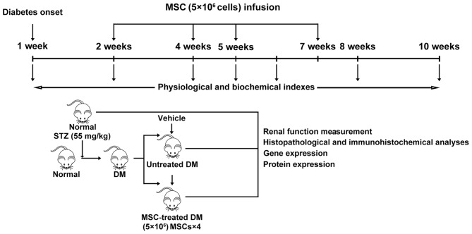
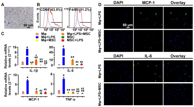
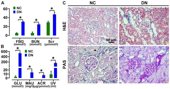
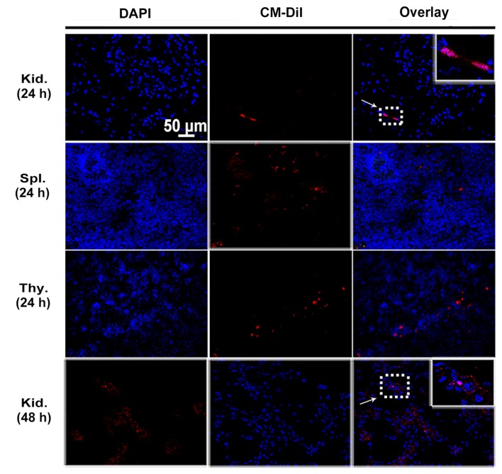
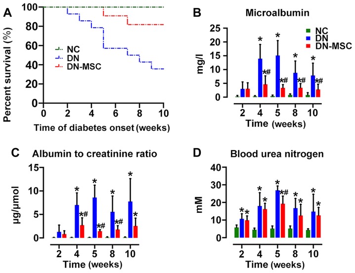
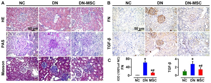
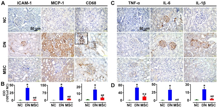
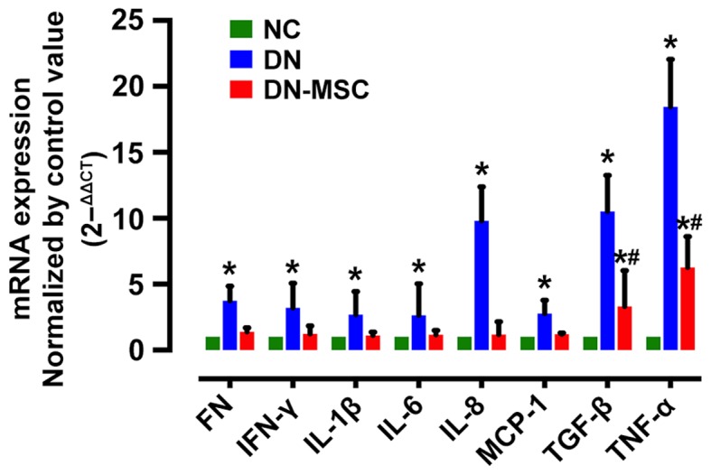
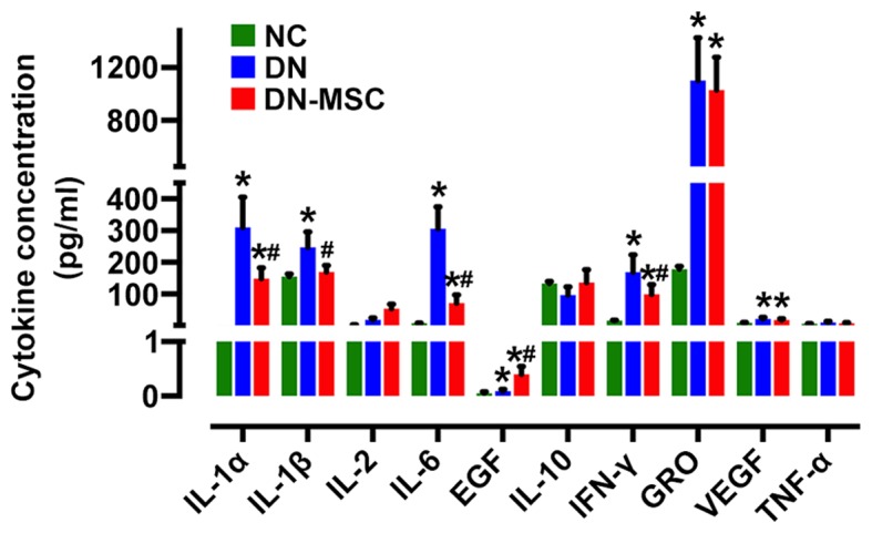
Diabetic nephropathy (DN) is a major complication of diabetes and represents the leading cause of end-stage renal disease. Mesenchymal stem cell (MSC) treatment has been demonstrated to be effective in DN models by reducing albuminuria and attenuating glomerular injury; however, limited in-depth understanding of the underlying mechanism and a lack of clinical trials hinders its clinical use. Additionally, most of these experimental studies were conducted on the advanced stage of nephropathy, which is difficult to reverse and consequently showed limited therapeutic efficacy. We sought to evaluate whether early intervention by MSCs has the potential to prevent DN onset and progression as well as protect kidney function when intravenously administered to rats with diabetes. Diabetes was induced in adult male SD rats by streptozotocin (STZ) injection (55 mg/kg, i.p.). The diabetic rats were injected with or without bone marrow-derived MSCs (5x10⁶ per rat), via tail vein at 2, 4, 5 and 7 weeks after diabetes onset. Fasting blood glucose (FBG), blood urea nitrogen (BUN) and serum creatinine (Scr) levels in serum samples and glycosuria (GLU), microalbumin (MAU), and albumin to creatinine ratio (ACR) in urine samples were determined. Renal pathology and immunohistochemistry (IHC) for CD68, MCP-1, fibronectin (FN), transforming growth factor-β (TGF-β) and pro-inflammatory cytokines were also performed. Expression levels of the above factors as well as interleukin-10 (IL-10), and epidermal growth factor (EGF) were assessed by qPCR and multiplex bead-based suspension array system, respectively. Additionally, MSC tracing in vivo was performed. Ex vivo, peritoneal macrophages were co-cultured with MSCs, and expression of inflammatory cytokines was detected as well. MSC treatment profoundly suppressed renal macrophage infiltration and inflammatory cytokine secretion in diabetic rats, resulting in prominently improved kidney histology, systemic homeostasis, and animal survival, although no significant effect on hyperglycemia was observed. Engrafted MSCs were primarily localized in deteriorated areas of the kidney and immune organs 48 h after infusion. MSC treatment upregulated serum anti-inflammatory cytokines IL-10 and EGF. Ex vivo, MSCs inhibited lipopolysaccharide (LPS)-stimulated rat peritoneal macrophage activation via the downregulation of inflammatory-related cytokines such as IL-6, MCP-1, tumor necrosis factor-α (TNF-α) and IL-1β. Our results demonstrated that early intervention with MSCs prevented renal injury via immune regulation in diabetic rats, which restored the homeostasis of the immune microenvironment, contributing to the prevention of kidney dysfunction and glomerulosclerosis.
糖尿病肾病(DN)是糖尿病的主要并发症,也是终末期肾病的主要病因。间充质干细胞(MSC)治疗已被证明可通过减少白蛋白尿和减轻肾小球损伤在 DN 模型中有效,然而,对其潜在机制的理解有限以及缺乏临床试验阻碍了其临床应用。此外,这些实验研究大多在肾病的晚期进行,此时肾病难以逆转,因此疗效有限。我们旨在评估间充质干细胞(MSC)早期干预是否有可能预防糖尿病大鼠的糖尿病发病和进展,并在静脉注射时保护肾功能。通过链脲佐菌素(STZ)(55mg/kg,ip)注射诱导成年雄性 SD 大鼠糖尿病。在糖尿病发病后 2、4、5 和 7 周,通过尾静脉向糖尿病大鼠注射或不注射骨髓来源的间充质干细胞(每只大鼠 5x10⁶)。测定血清样本中的空腹血糖(FBG)、血尿素氮(BUN)和血清肌酐(Scr)水平以及尿液样本中的尿糖(GLU)、微量白蛋白(MAU)和白蛋白/肌酐比(ACR)。还进行了肾组织病理学和免疫组化(IHC)检测 CD68、MCP-1、纤维连接蛋白(FN)、转化生长因子-β(TGF-β)和促炎细胞因子。通过 qPCR 和多因子珠悬浮阵列系统分别评估上述因子以及白细胞介素-10(IL-10)和表皮生长因子(EGF)的表达水平。此外,还进行了 MSC 的体内示踪。在体外,将腹腔巨噬细胞与 MSC 共培养,并检测炎性细胞因子的表达。MSC 治疗显著抑制了糖尿病大鼠肾巨噬细胞浸润和炎性细胞因子的分泌,从而显著改善了肾脏组织学、全身内环境平衡和动物存活率,尽管对高血糖没有明显影响。输注后 48 小时,移植的 MSC 主要定位于肾脏和免疫器官的受损区域。MSC 治疗上调了血清抗炎细胞因子 IL-10 和 EGF。在体外,MSC 通过下调 LPS 刺激的大鼠腹腔巨噬细胞中的炎性相关细胞因子(如 IL-6、MCP-1、肿瘤坏死因子-α(TNF-α)和 IL-1β)来抑制其激活。我们的结果表明,MSC 早期干预通过免疫调节预防了糖尿病大鼠的肾损伤,恢复了免疫微环境的内环境平衡,有助于预防肾功能障碍和肾小球硬化。