Visual Function Core, National Eye Institute, National Institutes of Health, Bethesda, Maryland, United States.
Unit on Neuron-Glia Interactions in Retinal Disease, National Eye Institute, National Institutes of Health, Bethesda, Maryland, United States.
Invest Ophthalmol Vis Sci. 2018 Feb 1;59(2):1084-1094. doi: 10.1167/iovs.17-23011.
Using optical coherence tomography (OCT) to analyze the effects of light/dark adaptation in a mouse model of inherited photoreceptor degeneration (rd10), and to study dynamics of subretinal fluid during the progress of retinal degeneration.
rd10 and wild-type (WT) C57BL/6J mice were reared in cyclic light or darkness and imaged with Bioptigen UHR-OCT or Spectralis HRA+OCT after adaptation to either light or darkness.
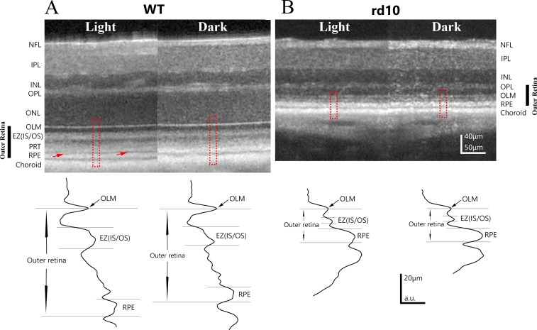
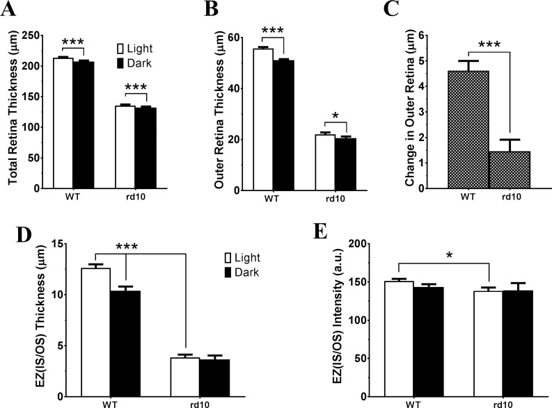
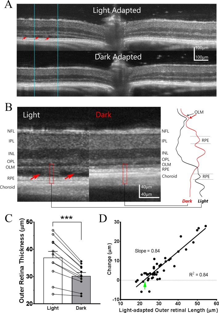
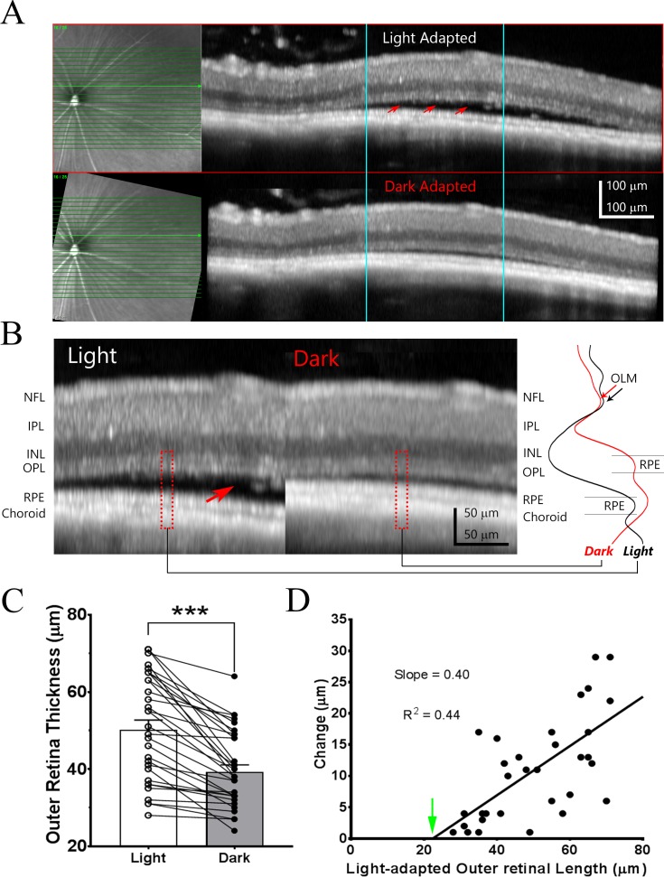
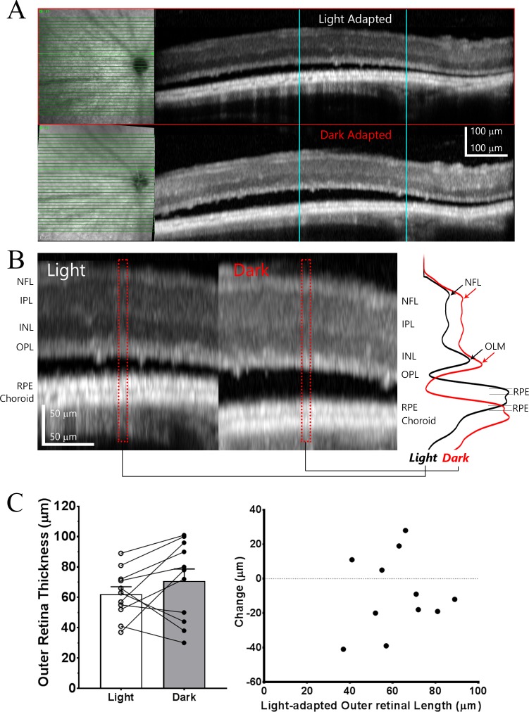
OCT images from rd10 mice were analyzed at three progressive stages of degeneration. After light-adaptation, stage I (postnatal age [P]26-29) eyes demonstrated no apparent subretinal fluid. At stage II (P32-38), subretinal fluid was present and restricted to parapapillary area, while at stage III (P44-45) extensive subretinal fluid was present across many retinal areas. Following overnight dark-adaptation, WT eyes showed a large reduction in outer retinal thickness (4.6 ± 1.4 μm, n = 16), whereas this change was significantly smaller in stage I rd10 eyes (1.5 ± 0.5 μm, n = 14). In stage II rd10 eyes, dark-adaptation significantly reduced the extent of subretinal fluid, with the amount of reduction correlating with the amount of fluid pre-existing in the light-adapted state. However, dark-adaptation did not significantly alter the amount of subretinal fluid observed in stage III rd10 mice. In addition, dark-rearing of rd10 mice from P6 to P30 slowed retinal degeneration.
Visual experience in the form of light/dark adaptation exerts a significant effect on outer retinal structure in the context of photoreceptor degeneration. This effect may arise from light-dependent alterations in fluid transport across the RPE monolayer, and promote photoreceptor survival as induced by dark-rearing.
利用光学相干断层扫描(OCT)分析遗传性光感受器变性(rd10)小鼠模型中光/暗适应的影响,并研究视网膜变性过程中视网膜下液的动力学。
rd10 和野生型(WT)C57BL/6J 小鼠在循环光照或黑暗中饲养,并在适应光照或黑暗后用 Bioptigen UHR-OCT 或 Spectralis HRA+OCT 进行成像。
在退行性变的三个渐进阶段分析了 rd10 小鼠的 OCT 图像。在光适应后,第 I 期(出生后年龄[P]26-29)眼睛没有明显的视网膜下液。在第 II 期(P32-38),视网膜下液存在且局限于旁乳头区域,而在第 III 期(P44-45),广泛的视网膜下液存在于许多视网膜区域。经过一夜的暗适应后,WT 眼睛的外视网膜厚度显著减小(4.6±1.4μm,n=16),而 I 期 rd10 眼睛的变化明显较小(1.5±0.5μm,n=14)。在 II 期 rd10 眼睛中,暗适应显著减少了视网膜下液的量,减少的量与光适应状态下预先存在的液体量相关。然而,暗适应并没有显著改变 III 期 rd10 小鼠观察到的视网膜下液量。此外,从 P6 到 P30 对 rd10 小鼠进行暗饲养可减缓视网膜变性。
光感受器变性背景下,光/暗适应的视觉体验对外层视网膜结构有显著影响。这种影响可能源于 RPE 单层中流体转运的光依赖性改变,并通过暗饲养诱导光感受器存活。