• 文献检索
  • 文档翻译
  • 深度研究
  • 学术资讯
  • Suppr Zotero 插件Zotero 插件
  • 邀请有礼
  • 套餐&价格
  • 历史记录
应用&插件
Suppr Zotero 插件Zotero 插件浏览器插件Mac 客户端Windows 客户端微信小程序
定价
高级版会员购买积分包购买API积分包
服务
文献检索文档翻译深度研究API 文档MCP 服务
关于我们
关于 Suppr公司介绍联系我们用户协议隐私条款
关注我们

Suppr 超能文献

核心技术专利:CN118964589B侵权必究
粤ICP备2023148730 号-1Suppr @ 2026

文献检索

告别复杂PubMed语法,用中文像聊天一样搜索,搜遍4000万医学文献。AI智能推荐,让科研检索更轻松。

立即免费搜索

文件翻译

保留排版,准确专业,支持PDF/Word/PPT等文件格式,支持 12+语言互译。

免费翻译文档

深度研究

AI帮你快速写综述,25分钟生成高质量综述,智能提取关键信息,辅助科研写作。

立即免费体验

衰老标志物在个体人类供体体内的体外和原位情况是否相关?

Do senescence markers correlate in vitro and in situ within individual human donors?

作者信息

Waaijer Mariëtte E C, Gunn David A, van Heemst Diana, Slagboom P Eline, Sedivy John M, Dirks Roeland W, Tanke Hans J, Westendorp Rudi G J, Maier Andrea B

机构信息

Department of Gerontology and Geriatrics, Leiden University Medical Center, 2300 RC Leiden, the Netherlands.

Unilever Discover, Colworth Science Park, Sharnbrook, Bedfordshire MK44 1LQ, UK.

出版信息

Aging (Albany NY). 2018 Feb 28;10(2):278-289. doi: 10.18632/aging.101389.

DOI:10.18632/aging.101389
PMID:29500330
原文链接:https://pmc.ncbi.nlm.nih.gov/articles/PMC5842854/
Abstract

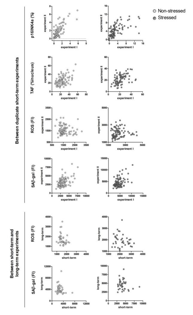
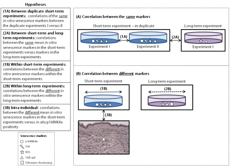
Little is known on how well senescence markers and correlate within individual donors. We studied correlations between the same and different markers. Furthermore, we tested correlations between markers with p16INK4a positivity.From 100 donors (20-91 years), cultured dermal fibroblasts were assessed for reactive oxygen species (ROS), telomere-associated foci (TAF), p16INK4a and senescence-associated β-gal (SAβ-gal), with/ without 0.6 µM rotenone for 3 days (short-term). In fibroblasts from 40 donors, telomere shortening, ROS and SAβ-gal were additionally assessed, with/ without 20 nM rotenone for 7 weeks (long-term). In skin from 52 donors, the number of p16INK4a positive dermal cells was assessed .More than half of the correlations of the same senescence markers between duplicate experiments and between short-term versus long-term experiments were significant. Half of the different senescence marker correlations were significant within the short-term and within the long-term experiments. The different senescence markers were not significantly correlated intra-individually with p16INK4a positivity.
In conclusion, the same and different senescence markers are frequently correlated significantly within and between experiments, but senescence markers are not correlated with p16INK4a positivity .

摘要

关于衰老标志物在个体供体中的相关性如何,目前所知甚少。我们研究了相同和不同标志物之间的相关性。此外,我们还测试了与p16INK4a阳性相关的标志物之间的相关性。从100名供体(20 - 91岁)中获取培养的真皮成纤维细胞,评估其活性氧(ROS)、端粒相关灶(TAF)、p16INK4a和衰老相关β - 半乳糖苷酶(SAβ - gal),在有/无0.6 μM鱼藤酮的情况下培养3天(短期)。在40名供体的成纤维细胞中,另外评估了端粒缩短、ROS和SAβ - gal,在有/无20 nM鱼藤酮的情况下培养7周(长期)。在52名供体的皮肤中,评估了p16INK4a阳性真皮细胞的数量。重复实验之间以及短期与长期实验之间,超过一半的相同衰老标志物的相关性是显著的。在短期和长期实验中,不同衰老标志物之间一半的相关性是显著的。不同的衰老标志物在个体内与p16INK4a阳性无显著相关性。总之,相同和不同的衰老标志物在实验内和实验间经常显著相关,但衰老标志物与p16INK4a阳性不相关。

https://cdn.ncbi.nlm.nih.gov/pmc/blobs/607c/5842854/a0bef28512da/aging-10-101389-g003.jpg
https://cdn.ncbi.nlm.nih.gov/pmc/blobs/607c/5842854/959c8d87025f/aging-10-101389-g001.jpg
https://cdn.ncbi.nlm.nih.gov/pmc/blobs/607c/5842854/479b2863fb93/aging-10-101389-g002.jpg
https://cdn.ncbi.nlm.nih.gov/pmc/blobs/607c/5842854/a0bef28512da/aging-10-101389-g003.jpg
https://cdn.ncbi.nlm.nih.gov/pmc/blobs/607c/5842854/959c8d87025f/aging-10-101389-g001.jpg
https://cdn.ncbi.nlm.nih.gov/pmc/blobs/607c/5842854/479b2863fb93/aging-10-101389-g002.jpg
https://cdn.ncbi.nlm.nih.gov/pmc/blobs/607c/5842854/a0bef28512da/aging-10-101389-g003.jpg

相似文献

1
Do senescence markers correlate in vitro and in situ within individual human donors?衰老标志物在个体人类供体体内的体外和原位情况是否相关?
Aging (Albany NY). 2018 Feb 28;10(2):278-289. doi: 10.18632/aging.101389.
2
[Expression of p16INK4a in nucleus pulposus and its effect on degenerated intervertebral discs].[P16INK4a在髓核中的表达及其对退变椎间盘的影响]
Zhongguo Xiu Fu Chong Jian Wai Ke Za Zhi. 2014 Dec;28(12):1514-8.
3
Aging is associated with decreased maximal life span and accelerated senescence of bone marrow stromal cells.衰老与最大寿命缩短和骨髓基质细胞衰老加速有关。
Bone. 2003 Dec;33(6):919-26. doi: 10.1016/j.bone.2003.07.005.
4
Telomere Homeostasis and Senescence Markers Are Differently Expressed in Placentas From Pregnancies With Early- Versus Late-Onset Preeclampsia.端粒稳态和衰老标志物在早发型与晚发型子痫前期胎盘组织中的表达不同。
Reprod Sci. 2019 Sep;26(9):1203-1209. doi: 10.1177/1933719118811644. Epub 2018 Nov 25.
5
Accelerated senescence in the kidneys of patients with type 2 diabetic nephropathy.2型糖尿病肾病患者肾脏的加速衰老
Am J Physiol Renal Physiol. 2008 Nov;295(5):F1563-73. doi: 10.1152/ajprenal.90302.2008. Epub 2008 Sep 3.
6
Markers of cellular senescence are elevated in murine blastocysts cultured in vitro: molecular consequences of culture in atmospheric oxygen.体外培养的小鼠囊胚中细胞衰老标志物升高:在大气氧环境中培养的分子后果。
J Assist Reprod Genet. 2014 Oct;31(10):1259-67. doi: 10.1007/s10815-014-0299-8. Epub 2014 Aug 10.
7
Long-term exposure to TNF-α leads human skin fibroblasts to a p38 MAPK- and ROS-mediated premature senescence.长期暴露于 TNF-α 会导致人皮肤成纤维细胞发生 p38 MAPK 和 ROS 介导的过早衰老。
Biogerontology. 2018 Jul;19(3-4):237-249. doi: 10.1007/s10522-018-9753-9. Epub 2018 Mar 26.
8
In vitro lifespan and senescence mechanisms of human nucleus pulposus chondrocytes.人髓核软骨细胞的体外寿命和衰老机制
Spine J. 2014 Mar 1;14(3):499-504. doi: 10.1016/j.spinee.2013.06.099. Epub 2013 Dec 15.
9
Vulnerability to oxidative stress and different patterns of senescence in human peritoneal mesothelial cell strains.人腹膜间皮细胞株对氧化应激的易感性及不同的衰老模式
Am J Physiol Regul Integr Comp Physiol. 2009 Feb;296(2):R374-82. doi: 10.1152/ajpregu.90451.2008. Epub 2008 Nov 26.
10
Accelerated cellular senescence in degenerate intervertebral discs: a possible role in the pathogenesis of intervertebral disc degeneration.退变椎间盘细胞的加速衰老:在椎间盘退变发病机制中的可能作用。
Arthritis Res Ther. 2007;9(3):R45. doi: 10.1186/ar2198.

引用本文的文献

1
A Functional Skincare Formulation Mixed With Retinyl Propionate, Hydroxypinacolone Retinoate, and Vitamin C on Antiaging and Whitening Han Women in Shanghai, China.一种含有丙酸视黄酯、羟基频哪酮视黄酸酯和维生素C的功能性护肤配方对中国上海汉族女性的抗衰及美白作用
J Cosmet Dermatol. 2025 Mar;24(3):e16747. doi: 10.1111/jocd.16747.
2
Cell Senescence-Independent Changes of Human Skin Fibroblasts with Age.人皮肤成纤维细胞衰老非依赖性的增龄变化。
Cells. 2024 Apr 9;13(8):659. doi: 10.3390/cells13080659.
3
Cellular Senescence in Human Skin Aging: Leveraging Senotherapeutics.

本文引用的文献

1
DNA damage markers in dermal fibroblasts in vitro reflect chronological donor age.体外培养的真皮成纤维细胞中的DNA损伤标志物反映了供体的实际年龄。
Aging (Albany NY). 2016 Jan;8(1):147-57. doi: 10.18632/aging.100890.
2
Forging a signature of in vivo senescence.伪造体内衰老的签名。
Nat Rev Cancer. 2015 Jul;15(7):397-408. doi: 10.1038/nrc3960.
3
Long-term Neuroglial Cocultures as a Brain Aging Model: Hallmarks of Senescence, MicroRNA Expression Profiles, and Comparison With In Vivo Models.作为脑衰老模型的长期神经胶质细胞共培养:衰老特征、微小RNA表达谱以及与体内模型的比较
人类皮肤衰老中的细胞衰老:利用衰老治疗学。
Gerontology. 2024;70(1):7-14. doi: 10.1159/000534756. Epub 2023 Oct 25.
4
Skin Aging in Long-Lived Naked Mole-Rats Is Accompanied by Increased Expression of Longevity-Associated and Tumor Suppressor Genes.长寿裸鼹鼠的皮肤衰老伴随着长寿相关和肿瘤抑制基因表达的增加。
J Invest Dermatol. 2022 Nov;142(11):2853-2863.e4. doi: 10.1016/j.jid.2022.04.028. Epub 2022 Jun 9.
5
Age-Related Changes in the Fibroblastic Differon of the Dermis: Role in Skin Aging.真皮成纤维细胞分化的年龄相关性变化:在皮肤衰老中的作用。
Int J Mol Sci. 2022 May 30;23(11):6135. doi: 10.3390/ijms23116135.
6
Skin Aging, Cellular Senescence and Natural Polyphenols.皮肤衰老、细胞衰老与天然多酚
Int J Mol Sci. 2021 Nov 23;22(23):12641. doi: 10.3390/ijms222312641.
7
Cellular Senescence and Inflammaging in the Skin Microenvironment.皮肤微环境中的细胞衰老和炎症老化。
Int J Mol Sci. 2021 Apr 8;22(8):3849. doi: 10.3390/ijms22083849.
8
LPS-induced premature osteocyte senescence: Implications in inflammatory alveolar bone loss and periodontal disease pathogenesis.脂多糖诱导的成骨细胞过早衰老:对炎症性牙槽骨丧失和牙周病发病机制的影响
Bone. 2020 Mar;132:115220. doi: 10.1016/j.bone.2019.115220. Epub 2020 Jan 2.
9
LncRNAs Regulatory Networks in Cellular Senescence.lncRNAs 调控细胞衰老的网络。
Int J Mol Sci. 2019 May 28;20(11):2615. doi: 10.3390/ijms20112615.
10
Biomarkers of Cellular Senescence and Skin Aging.细胞衰老与皮肤老化的生物标志物
Front Genet. 2018 Aug 23;9:247. doi: 10.3389/fgene.2018.00247. eCollection 2018.
J Gerontol A Biol Sci Med Sci. 2016 Jan;71(1):50-60. doi: 10.1093/gerona/glu231. Epub 2015 Jan 7.
4
Physiological and pathological consequences of cellular senescence.细胞衰老的生理和病理后果。
Cell Mol Life Sci. 2014 Nov;71(22):4373-86. doi: 10.1007/s00018-014-1691-3. Epub 2014 Jul 31.
5
The hallmarks of fibroblast ageing.成纤维细胞衰老的特征。
Mech Ageing Dev. 2014 Jun;138:26-44. doi: 10.1016/j.mad.2014.03.004. Epub 2014 Mar 29.
6
In vivo and in vitro analysis of age-associated changes and somatic cellular senescence in renal epithelial cells.肾上皮细胞中与年龄相关变化及体细胞衰老的体内和体外分析
PLoS One. 2014 Feb 4;9(2):e88071. doi: 10.1371/journal.pone.0088071. eCollection 2014.
7
p16INK4a reporter mice reveal age-promoting effects of environmental toxicants.p16INK4a 报告基因小鼠揭示环境毒物的促衰老作用。
J Clin Invest. 2014 Jan;124(1):169-73. doi: 10.1172/JCI70960. Epub 2013 Dec 16.
8
Programmed cell senescence during mammalian embryonic development.哺乳动物胚胎发育过程中的程序性细胞衰老。
Cell. 2013 Nov 21;155(5):1104-18. doi: 10.1016/j.cell.2013.10.019. Epub 2013 Nov 14.
9
Senescence is a developmental mechanism that contributes to embryonic growth and patterning.衰老(Senescence)是一种发育机制,有助于胚胎的生长和模式形成。
Cell. 2013 Nov 21;155(5):1119-30. doi: 10.1016/j.cell.2013.10.041. Epub 2013 Nov 14.
10
Monitoring tumorigenesis and senescence in vivo with a p16(INK4a)-luciferase model.利用 p16(INK4a)-荧光素酶模型在体内监测肿瘤发生和衰老。
Cell. 2013 Jan 17;152(1-2):340-51. doi: 10.1016/j.cell.2012.12.010.