• 文献检索
  • 文档翻译
  • 深度研究
  • 学术资讯
  • Suppr Zotero 插件Zotero 插件
  • 邀请有礼
  • 套餐&价格
  • 历史记录
应用&插件
Suppr Zotero 插件Zotero 插件浏览器插件Mac 客户端Windows 客户端微信小程序
定价
高级版会员购买积分包购买API积分包
服务
文献检索文档翻译深度研究API 文档MCP 服务
关于我们
关于 Suppr公司介绍联系我们用户协议隐私条款
关注我们

Suppr 超能文献

核心技术专利:CN118964589B侵权必究
粤ICP备2023148730 号-1Suppr @ 2026

文献检索

告别复杂PubMed语法,用中文像聊天一样搜索,搜遍4000万医学文献。AI智能推荐,让科研检索更轻松。

立即免费搜索

文件翻译

保留排版,准确专业,支持PDF/Word/PPT等文件格式,支持 12+语言互译。

免费翻译文档

深度研究

AI帮你快速写综述,25分钟生成高质量综述,智能提取关键信息,辅助科研写作。

立即免费体验

减少周细胞衍生的瘢痕形成可促进脊髓损伤后的恢复。

Reducing Pericyte-Derived Scarring Promotes Recovery after Spinal Cord Injury.

机构信息

Department of Cell and Molecular Biology, Karolinska Institutet, SE-171 77 Stockholm, Sweden.

Department of Neuroscience, Karolinska Institutet, SE-171 77 Stockholm, Sweden.

出版信息

Cell. 2018 Mar 22;173(1):153-165.e22. doi: 10.1016/j.cell.2018.02.004. Epub 2018 Mar 1.

DOI:10.1016/j.cell.2018.02.004
PMID:29502968
原文链接:https://pmc.ncbi.nlm.nih.gov/articles/PMC5871719/
Abstract

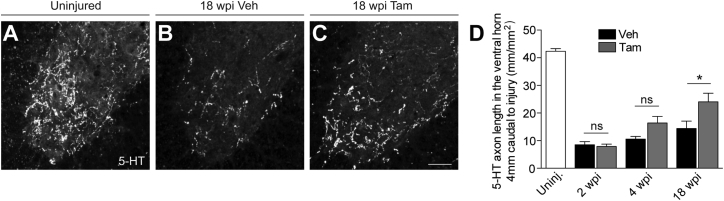
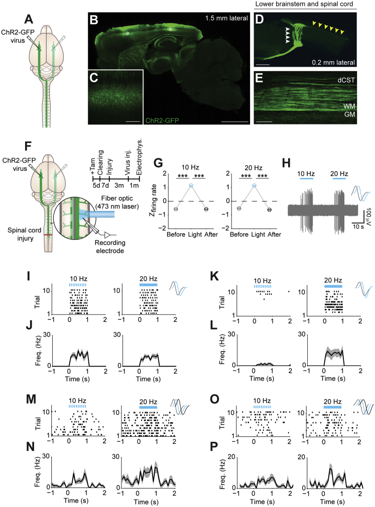
CNS injury often severs axons. Scar tissue that forms locally at the lesion site is thought to block axonal regeneration, resulting in permanent functional deficits. We report that inhibiting the generation of progeny by a subclass of pericytes led to decreased fibrosis and extracellular matrix deposition after spinal cord injury in mice. Regeneration of raphespinal and corticospinal tract axons was enhanced and sensorimotor function recovery improved following spinal cord injury in animals with attenuated pericyte-derived scarring. Using optogenetic stimulation, we demonstrate that regenerated corticospinal tract axons integrated into the local spinal cord circuitry below the lesion site. The number of regenerated axons correlated with improved sensorimotor function recovery. In conclusion, attenuation of pericyte-derived fibrosis represents a promising therapeutic approach to facilitate recovery following CNS injury.

摘要

中枢神经系统损伤常导致轴突断裂。损伤部位局部形成的疤痕组织被认为会阻碍轴突再生,导致永久性功能缺陷。我们报告称,抑制一小部分周细胞的后代生成会导致小鼠脊髓损伤后纤维化和细胞外基质沉积减少。损伤后,减少周细胞来源的疤痕形成会增强中缝核脊髓束和皮质脊髓束轴突的再生,改善感觉运动功能恢复。通过光遗传刺激,我们证明再生的皮质脊髓束轴突整合到损伤部位下方的局部脊髓回路中。再生轴突的数量与感觉运动功能恢复的改善相关。总之,减少周细胞来源的纤维化是一种很有前途的治疗方法,可以促进中枢神经系统损伤后的恢复。

https://cdn.ncbi.nlm.nih.gov/pmc/blobs/82aa/5871719/5ad7d26f6ed6/figs7.jpg
https://cdn.ncbi.nlm.nih.gov/pmc/blobs/82aa/5871719/6bf6dcb8ed0a/fx1.jpg
https://cdn.ncbi.nlm.nih.gov/pmc/blobs/82aa/5871719/e7400aee12db/gr1.jpg
https://cdn.ncbi.nlm.nih.gov/pmc/blobs/82aa/5871719/8bb42555bc76/figs1.jpg
https://cdn.ncbi.nlm.nih.gov/pmc/blobs/82aa/5871719/b2c05a3d6060/figs2.jpg
https://cdn.ncbi.nlm.nih.gov/pmc/blobs/82aa/5871719/a07a76810f36/figs3.jpg
https://cdn.ncbi.nlm.nih.gov/pmc/blobs/82aa/5871719/0c81457efc51/gr2.jpg
https://cdn.ncbi.nlm.nih.gov/pmc/blobs/82aa/5871719/3cdfe2106902/gr3.jpg
https://cdn.ncbi.nlm.nih.gov/pmc/blobs/82aa/5871719/a02b3406c102/figs4.jpg
https://cdn.ncbi.nlm.nih.gov/pmc/blobs/82aa/5871719/04005a37e08d/gr4.jpg
https://cdn.ncbi.nlm.nih.gov/pmc/blobs/82aa/5871719/89cd3e7c07e0/gr5.jpg
https://cdn.ncbi.nlm.nih.gov/pmc/blobs/82aa/5871719/d4cbe2c4d3fc/figs5.jpg
https://cdn.ncbi.nlm.nih.gov/pmc/blobs/82aa/5871719/442f639a35e1/figs6.jpg
https://cdn.ncbi.nlm.nih.gov/pmc/blobs/82aa/5871719/14afed284b55/gr6.jpg
https://cdn.ncbi.nlm.nih.gov/pmc/blobs/82aa/5871719/68db4f2feb03/gr7.jpg
https://cdn.ncbi.nlm.nih.gov/pmc/blobs/82aa/5871719/5ad7d26f6ed6/figs7.jpg

相似文献

1
Reducing Pericyte-Derived Scarring Promotes Recovery after Spinal Cord Injury.减少周细胞衍生的瘢痕形成可促进脊髓损伤后的恢复。
Cell. 2018 Mar 22;173(1):153-165.e22. doi: 10.1016/j.cell.2018.02.004. Epub 2018 Mar 1.
2
A switch in pdgfrb cell-derived ECM composition prevents inhibitory scarring and promotes axon regeneration in the zebrafish spinal cord.PDGFRB 细胞衍生细胞外基质组成的转变可防止抑制性瘢痕形成,并促进斑马鱼脊髓中的轴突再生。
Dev Cell. 2021 Feb 22;56(4):509-524.e9. doi: 10.1016/j.devcel.2020.12.009. Epub 2021 Jan 6.
3
Proliferating NG2-Cell-Dependent Angiogenesis and Scar Formation Alter Axon Growth and Functional Recovery After Spinal Cord Injury in Mice.NG2 细胞依赖性血管生成和瘢痕形成促进小鼠脊髓损伤后的轴突生长和功能恢复。
J Neurosci. 2018 Feb 7;38(6):1366-1382. doi: 10.1523/JNEUROSCI.3953-16.2017. Epub 2017 Dec 26.
4
Pten Deletion Promotes Regrowth of Corticospinal Tract Axons 1 Year after Spinal Cord Injury.脊髓损伤1年后,PTEN缺失促进皮质脊髓束轴突再生。
J Neurosci. 2015 Jul 1;35(26):9754-63. doi: 10.1523/JNEUROSCI.3637-14.2015.
5
Pericyte-derived fibrotic scarring is conserved across diverse central nervous system lesions.周细胞衍生的纤维瘢痕在不同的中枢神经系统病变中是保守存在的。
Nat Commun. 2021 Sep 17;12(1):5501. doi: 10.1038/s41467-021-25585-5.
6
Suppression of fibrous scarring in spinal cord injury of rat promotes long-distance regeneration of corticospinal tract axons, rescue of primary motoneurons in somatosensory cortex and significant functional recovery.抑制大鼠脊髓损伤中的纤维瘢痕形成可促进皮质脊髓束轴突的长距离再生、挽救体感皮层中的初级运动神经元并实现显著的功能恢复。
Eur J Neurosci. 2005 Dec;22(12):3047-58. doi: 10.1111/j.1460-9568.2005.04495.x.
7
The TGFβ1/SMADs/Snail1 signaling axis mediates pericyte-derived fibrous scar formation after spinal cord injury.TGFβ1/SMADs/Snail1 信号轴介导脊髓损伤后周细胞来源的纤维瘢痕形成。
Int Immunopharmacol. 2024 Feb 15;128:111482. doi: 10.1016/j.intimp.2023.111482. Epub 2024 Jan 18.
8
Fibrotic scarring following lesions to the central nervous system.中枢神经系统损伤后的纤维化瘢痕形成。
Matrix Biol. 2018 Aug;68-69:561-570. doi: 10.1016/j.matbio.2018.02.009. Epub 2018 Feb 9.
9
Optogenetic Interrogation of Functional Synapse Formation by Corticospinal Tract Axons in the Injured Spinal Cord.脊髓损伤后皮质脊髓束轴突功能性突触形成的光遗传学研究
J Neurosci. 2016 May 25;36(21):5877-90. doi: 10.1523/JNEUROSCI.4203-15.2016.
10
Pharmacological Suppression of CNS Scarring by Deferoxamine Reduces Lesion Volume and Increases Regeneration in an In Vitro Model for Astroglial-Fibrotic Scarring and in Rat Spinal Cord Injury In Vivo.去铁胺对中枢神经系统瘢痕形成的药理学抑制作用可减小体外星形胶质细胞纤维化瘢痕模型及大鼠体内脊髓损伤模型中的损伤体积并促进再生。
PLoS One. 2015 Jul 29;10(7):e0134371. doi: 10.1371/journal.pone.0134371. eCollection 2015.

引用本文的文献

1
Augmenting AMPA receptor signaling after spinal cord injury increases ependymal-derived neural stem/progenitor cell migration and promotes functional recovery.脊髓损伤后增强AMPA受体信号可增加室管膜来源的神经干细胞/祖细胞迁移并促进功能恢复。
Nat Neurosci. 2025 Sep 15. doi: 10.1038/s41593-025-02044-8.
2
Cutting-edge technologies in neural regeneration.神经再生领域的前沿技术。
Cell Regen. 2025 Sep 5;14(1):38. doi: 10.1186/s13619-025-00260-y.
3
Dynamic fibroblast-immune interactions shape recovery after brain injury.动态的成纤维细胞 - 免疫相互作用塑造脑损伤后的恢复过程。

本文引用的文献

1
Myelinogenic Plasticity of Oligodendrocyte Precursor Cells following Spinal Cord Contusion Injury.脊髓挫伤损伤后少突胶质前体细胞的髓鞘形成可塑性
J Neurosci. 2017 Sep 6;37(36):8635-8654. doi: 10.1523/JNEUROSCI.2409-16.2017. Epub 2017 Jul 31.
2
Interaction of reactive astrocytes with type I collagen induces astrocytic scar formation through the integrin-N-cadherin pathway after spinal cord injury.反应性星形胶质细胞与 I 型胶原的相互作用通过整合素-N-钙黏蛋白通路诱导脊髓损伤后星形胶质细胞瘢痕形成。
Nat Med. 2017 Jul;23(7):818-828. doi: 10.1038/nm.4354. Epub 2017 Jun 19.
3
Anatomical and Molecular Properties of Long Descending Propriospinal Neurons in Mice.
Nature. 2025 Sep 3. doi: 10.1038/s41586-025-09449-2.
4
Enhanced locomotor recovery in mice lacking GlcNAc6ST1 and GlcNAc6ST4 following spinal cord injury.脊髓损伤后缺乏GlcNAc6ST1和GlcNAc6ST4的小鼠运动功能恢复增强。
Life Sci Alliance. 2025 Aug 28;8(11). doi: 10.26508/lsa.202503469. Print 2025 Nov.
5
Single-Cell RNA Sequencing Reveals Pericytes Acquire a Fibrotic Phenotype and Promote Mesenteric Adipose Tissue Fibrosis in Crohn's Disease.单细胞RNA测序揭示周细胞获得纤维化表型并促进克罗恩病患者肠系膜脂肪组织纤维化
J Inflamm Res. 2025 Aug 14;18:11055-11071. doi: 10.2147/JIR.S528171. eCollection 2025.
6
F-box/LRR-repeat protein 12 reorchestrated microglia to inhibit scarring and achieve adult spinal cord injury repair.F-box/富含亮氨酸重复序列蛋白12重新编排小胶质细胞以抑制瘢痕形成并实现成年脊髓损伤修复。
Signal Transduct Target Ther. 2025 Aug 20;10(1):259. doi: 10.1038/s41392-025-02354-0.
7
Characteristics of Scar Formation After Intracerebral Hemorrhage in Aged Rats: Effects of Deferoxamine.老年大鼠脑出血后瘢痕形成的特征:去铁胺的作用
Cells. 2025 Jul 22;14(15):1127. doi: 10.3390/cells14151127.
8
Pericytes in tissue fibrosis.组织纤维化中的周细胞
Am J Physiol Cell Physiol. 2025 Sep 1;329(3):C868-C886. doi: 10.1152/ajpcell.00403.2025. Epub 2025 Aug 12.
9
The efficacy and pharmacological mechanisms of traditional Chinese medicine in the treatment of spinal cord injury.中药治疗脊髓损伤的疗效及药理机制
J Orthop Surg Res. 2025 Aug 8;20(1):748. doi: 10.1186/s13018-025-06129-9.
10
in vivo Reprogramming of NG2 Glia Improves Bladder Function After Spinal Cord Injury.脊髓损伤后,体内对少突胶质前体细胞进行重编程可改善膀胱功能。
bioRxiv. 2025 Jul 31:2025.07.28.667292. doi: 10.1101/2025.07.28.667292.
小鼠长下行脊髓 propriospinal 神经元的解剖学和分子特性
Front Neuroanat. 2017 Feb 6;11:5. doi: 10.3389/fnana.2017.00005. eCollection 2017.
4
Pericytes of Multiple Organs Do Not Behave as Mesenchymal Stem Cells In Vivo.多器官的周细胞在体内并非表现为间充质干细胞。
Cell Stem Cell. 2017 Mar 2;20(3):345-359.e5. doi: 10.1016/j.stem.2016.12.006. Epub 2017 Jan 19.
5
Understanding the NG2 Glial Scar after Spinal Cord Injury.了解脊髓损伤后的NG2胶质瘢痕。
Front Neurol. 2016 Nov 15;7:199. doi: 10.3389/fneur.2016.00199. eCollection 2016.
6
Analysis of the brain mural cell transcriptome.脑壁细胞转录组分析。
Sci Rep. 2016 Oct 11;6:35108. doi: 10.1038/srep35108.
7
The corticospinal tract: Evolution, development, and human disorders.皮质脊髓束:进化、发育及人类疾病
Dev Neurobiol. 2017 Jul;77(7):810-829. doi: 10.1002/dneu.22455. Epub 2016 Oct 14.
8
The glial scar is more than just astrocytes.神经胶质瘢痕不仅仅是星形胶质细胞。
Exp Neurol. 2016 Dec;286:147-149. doi: 10.1016/j.expneurol.2016.06.018. Epub 2016 Jun 18.
9
Optogenetic Interrogation of Functional Synapse Formation by Corticospinal Tract Axons in the Injured Spinal Cord.脊髓损伤后皮质脊髓束轴突功能性突触形成的光遗传学研究
J Neurosci. 2016 May 25;36(21):5877-90. doi: 10.1523/JNEUROSCI.4203-15.2016.
10
Astrocyte scar formation aids central nervous system axon regeneration.星形胶质细胞瘢痕形成有助于中枢神经系统轴突再生。
Nature. 2016 Apr 14;532(7598):195-200. doi: 10.1038/nature17623. Epub 2016 Mar 30.