Translational Neuropsychiatry Unit, Department of Clinical Medicine, Aarhus University, Risskov, Denmark.
Core Center for Molecular Morphology, Section for Stereology and Microscopy, Department of Clinical Medicine, Aarhus University, Aarhus, Denmark.
Int J Neuropsychopharmacol. 2018 Jun 1;21(6):603-615. doi: 10.1093/ijnp/pyy022.
Preclinical studies have indicated that antidepressant effect of vortioxetine involves increased synaptic plasticity and promotion of spine maturation. Mitochondria dysfunction may contribute to the pathophysiological basis of major depressive disorder. Taking into consideration that vortioxetine increases spine number and dendritic branching in hippocampus CA1 faster than fluoxetine, we hypothesize that new spines induced by vortioxetine can rapidly form functional synapses by mitochondrial support, accompanied by increased brain-derived neurotrophic factor signaling.
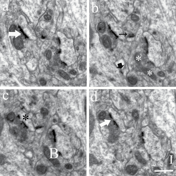
Rats were treated for 1 week with vortioxetine or fluoxetine at pharmacologically relevant doses. Number of synapses and mitochondria in hippocampus CA1 were quantified by electron microscopy. Brain-derived neurotrophic factor protein levels were visualized with immunohistochemistry. Gene and protein expression of synapse and mitochondria-related markers were investigated with real-time quantitative polymerase chain reaction and immunoblotting.
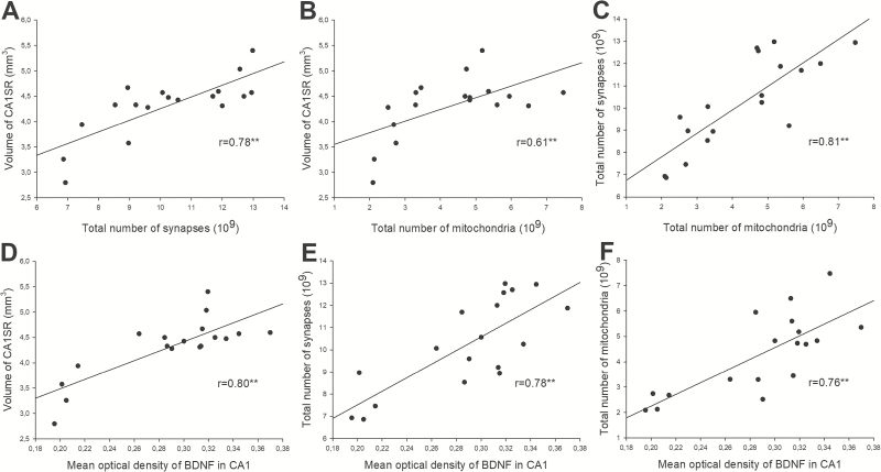
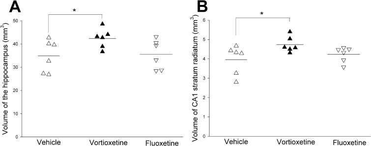
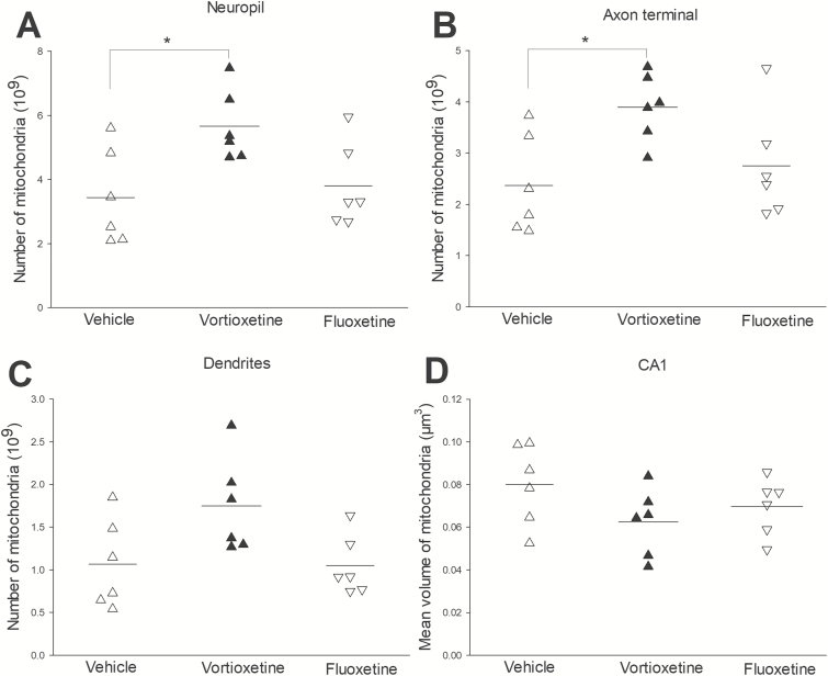
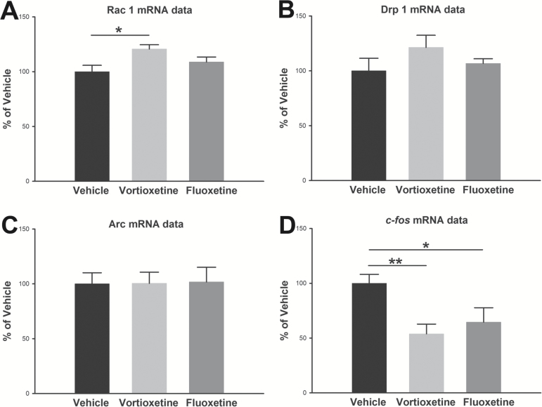
Vortioxetine increased number of synapses and mitochondria significantly, whereas fluoxetine had no effect after 1-week dosing. BDNF levels in hippocampus DG and CA1 were significantly higher after vortioxetine treatment. Gene expression levels of Rac1 after vortioxetine treatment were significantly increased. There was a tendency towards increased gene expression levels of Drp1 and protein levels of Rac1. However, both gene and protein levels of c-Fos were significantly decreased. Furthermore, there was a significant positive correlation between BDNF levels and mitochondria and synapse numbers.
Our results imply that mitochondria play a critical role in synaptic plasticity accompanied by increased BDNF levels. Rapid changes in BDNF levels and synaptic/mitochondria plasticity of hippocampus following vortioxetine compared with fluoxetine may be ascribed to vortioxetine's modulation of serotonin receptors.
临床前研究表明,文拉法辛具有抗抑郁作用,涉及增加突触可塑性和促进脊柱成熟。线粒体功能障碍可能是导致重度抑郁症病理生理基础的原因之一。考虑到文拉法辛比氟西汀更快地增加海马 CA1 中的棘突数量和树突分支,我们假设文拉法辛诱导的新棘突可以通过线粒体支持迅速形成功能性突触,伴随着脑源性神经营养因子信号的增加。
大鼠用文拉法辛或氟西汀以药理相关剂量治疗 1 周。用电子显微镜定量海马 CA1 中的突触和线粒体数量。用免疫组织化学法可视化脑源性神经营养因子蛋白水平。用实时定量聚合酶链反应和免疫印迹法研究突触和线粒体相关标志物的基因和蛋白表达。
文拉法辛显著增加突触和线粒体数量,而氟西汀在 1 周给药后无影响。文拉法辛治疗后海马 DG 和 CA1 中的 BDNF 水平显著升高。文拉法辛治疗后 Rac1 的基因表达水平显著增加。Drp1 的基因表达水平和 Rac1 的蛋白水平有增加的趋势。然而,c-Fos 的基因和蛋白水平均显著降低。此外,BDNF 水平与线粒体和突触数量之间存在显著的正相关。
我们的结果表明,线粒体在 BDNF 水平升高伴随的突触可塑性中起关键作用。与氟西汀相比,文拉法辛对海马体中 BDNF 水平和突触/线粒体可塑性的快速变化可能归因于文拉法辛对 5-羟色胺受体的调节。