• 文献检索
  • 文档翻译
  • 深度研究
  • 学术资讯
  • Suppr Zotero 插件Zotero 插件
  • 邀请有礼
  • 套餐&价格
  • 历史记录
应用&插件
Suppr Zotero 插件Zotero 插件浏览器插件Mac 客户端Windows 客户端微信小程序
定价
高级版会员购买积分包购买API积分包
服务
文献检索文档翻译深度研究API 文档MCP 服务
关于我们
关于 Suppr公司介绍联系我们用户协议隐私条款
关注我们

Suppr 超能文献

核心技术专利:CN118964589B侵权必究
粤ICP备2023148730 号-1Suppr @ 2026

文献检索

告别复杂PubMed语法,用中文像聊天一样搜索,搜遍4000万医学文献。AI智能推荐,让科研检索更轻松。

立即免费搜索

文件翻译

保留排版,准确专业,支持PDF/Word/PPT等文件格式,支持 12+语言互译。

免费翻译文档

深度研究

AI帮你快速写综述,25分钟生成高质量综述,智能提取关键信息,辅助科研写作。

立即免费体验

基于表观弥散系数图的全瘤体直方图分析鉴别胸腺癌与淋巴瘤。

A Whole-Tumor Histogram Analysis of Apparent Diffusion Coefficient Maps for Differentiating Thymic Carcinoma from Lymphoma.

机构信息

Department of Radiology, The First Affiliated Hospital of Nanjing Medical University, Nanjing 210000, China.

Department of Thoracic Surgery, The First Affiliated Hospital of Nanjing Medical University, Nanjing 210000, China.

出版信息

Korean J Radiol. 2018 Mar-Apr;19(2):358-365. doi: 10.3348/kjr.2018.19.2.358. Epub 2018 Feb 22.

DOI:10.3348/kjr.2018.19.2.358
PMID:29520195
原文链接:https://pmc.ncbi.nlm.nih.gov/articles/PMC5840066/
Abstract

OBJECTIVE

To assess the performance of a whole-tumor histogram analysis of apparent diffusion coefficient (ADC) maps in differentiating thymic carcinoma from lymphoma, and compare it with that of a commonly used hot-spot region-of-interest (ROI)-based ADC measurement.

MATERIALS AND METHODS

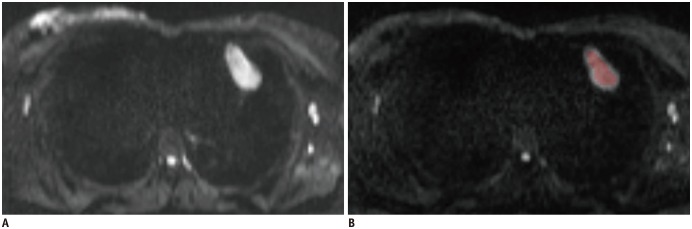
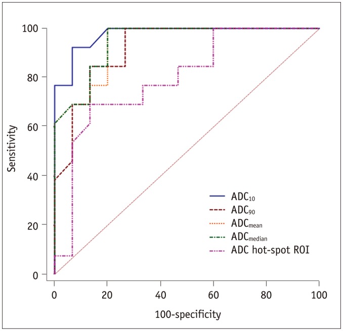
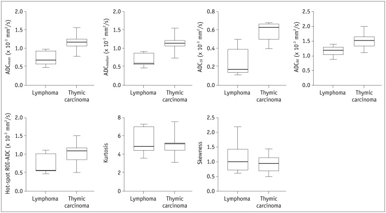
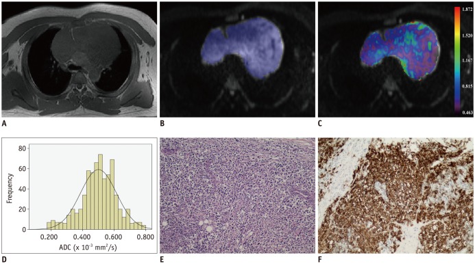
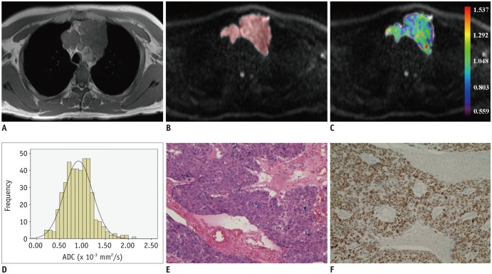
Diffusion weighted imaging data of 15 patients with thymic carcinoma and 13 patients with lymphoma were retrospectively collected and processed with a mono-exponential model. ADC measurements were performed by using a histogram-based and hot-spot-ROI-based approach. In the histogram-based approach, the following parameters were generated: mean ADC (ADC), median ADC (ADC), 10th and 90th percentile of ADC (ADC and ADC), kurtosis, and skewness. The difference in ADCs between thymic carcinoma and lymphoma was compared using a test. Receiver operating characteristic analyses were conducted to determine and compare the differentiating performance of ADCs.

RESULTS

Lymphoma demonstrated significantly lower ADC, ADC, ADC, ADC, and hot-spot-ROI-based mean ADC than those found in thymic carcinoma (all values < 0.05). There were no differences found in the kurtosis ( = 0.412) and skewness ( = 0.273). The ADC demonstrated optimal differentiating performance (cut-off value, 0.403 × 10 mm/s; area under the receiver operating characteristic curve [AUC], 0.977; sensitivity, 92.3%; specificity, 93.3%), followed by the ADC, ADC, ADC, and hot-spot-ROI-based mean ADC. The AUC of ADC was significantly higher than that of the hot spot ROI based ADC (0.977 vs. 0.797, = 0.036).

CONCLUSION

Compared with the commonly used hot spot ROI based ADC measurement, a histogram analysis of ADC maps can improve the differentiating performance between thymic carcinoma and lymphoma.

摘要

目的

评估表观扩散系数(ADC)图全瘤直方图分析在鉴别胸腺癌与淋巴瘤中的性能,并与常用的热点感兴趣区(ROI)-基于 ADC 测量进行比较。

材料与方法

回顾性收集 15 例胸腺癌和 13 例淋巴瘤患者的弥散加权成像数据,并采用单指数模型进行处理。采用直方图和热点 ROI 两种方法进行 ADC 测量。在直方图方法中,生成以下参数:平均 ADC(ADC)、中位数 ADC(ADC)、ADC 的第 10 百分位数和第 90 百分位数(ADC 和 ADC)、峰度和偏度。使用 t 检验比较胸腺癌和淋巴瘤之间 ADC 的差异。进行受试者工作特征分析以确定和比较 ADC 的鉴别性能。

结果

淋巴瘤的 ADC、ADC、ADC、ADC 和热点 ROI 平均 ADC 均显著低于胸腺癌(所有 P 值均<0.05)。峰度(=0.412)和偏度(=0.273)无差异。ADC 表现出最佳的鉴别性能(截断值,0.403×10 mm/s;受试者工作特征曲线下面积 [AUC],0.977;敏感性,92.3%;特异性,93.3%),其次是 ADC、ADC、ADC 和热点 ROI 平均 ADC。ADC 的 AUC 显著高于热点 ROI 基于 ADC(0.977 比 0.797,=0.036)。

结论

与常用的热点 ROI 基于 ADC 测量相比,ADC 图的直方图分析可以提高胸腺癌与淋巴瘤的鉴别性能。

https://cdn.ncbi.nlm.nih.gov/pmc/blobs/8514/5840066/64b35a713dde/kjr-19-358-g005.jpg
https://cdn.ncbi.nlm.nih.gov/pmc/blobs/8514/5840066/be837147b615/kjr-19-358-g001.jpg
https://cdn.ncbi.nlm.nih.gov/pmc/blobs/8514/5840066/958601d00231/kjr-19-358-g002.jpg
https://cdn.ncbi.nlm.nih.gov/pmc/blobs/8514/5840066/c77e5a37053f/kjr-19-358-g003.jpg
https://cdn.ncbi.nlm.nih.gov/pmc/blobs/8514/5840066/3ea5863a58c3/kjr-19-358-g004.jpg
https://cdn.ncbi.nlm.nih.gov/pmc/blobs/8514/5840066/64b35a713dde/kjr-19-358-g005.jpg

相似文献

1
A Whole-Tumor Histogram Analysis of Apparent Diffusion Coefficient Maps for Differentiating Thymic Carcinoma from Lymphoma.基于表观弥散系数图的全瘤体直方图分析鉴别胸腺癌与淋巴瘤。
Korean J Radiol. 2018 Mar-Apr;19(2):358-365. doi: 10.3348/kjr.2018.19.2.358. Epub 2018 Feb 22.
2
Histogram analysis of apparent diffusion coefficient maps for assessing thymic epithelial tumours: correlation with world health organization classification and clinical staging.表观扩散系数图的直方图分析用于评估胸腺上皮肿瘤:与世界卫生组织分类及临床分期的相关性
Br J Radiol. 2018 Apr;91(1084):20170580. doi: 10.1259/bjr.20170580. Epub 2018 Jan 10.
3
Radiological indeterminate vestibular schwannoma and meningioma in cerebellopontine angle area: differentiating using whole-tumor histogram analysis of apparent diffusion coefficient.桥小脑角区放射学表现不典型的前庭神经鞘瘤和脑膜瘤:利用表观扩散系数的全肿瘤直方图分析进行鉴别
Int J Neurosci. 2017 Feb;127(2):183-190. doi: 10.3109/00207454.2016.1164157. Epub 2016 Apr 6.
4
Histogram analysis of apparent diffusion coefficient maps for differentiating malignant from benign parotid gland tumors.用于区分腮腺良恶性肿瘤的表观扩散系数图的直方图分析
Eur Arch Otorhinolaryngol. 2018 Aug;275(8):2151-2157. doi: 10.1007/s00405-018-5052-y. Epub 2018 Jul 2.
5
Orbital Indeterminate Lesions in Adults: Combined Magnetic Resonance Morphometry and Histogram Analysis of Apparent Diffusion Coefficient Maps for Predicting Malignancy.成人眼眶不确定性病变:联合磁共振形态测量和表观扩散系数图的直方图分析预测恶性肿瘤
Acad Radiol. 2016 Feb;23(2):200-8. doi: 10.1016/j.acra.2015.10.015. Epub 2015 Nov 28.
6
Whole-tumor histogram analysis of apparent diffusion coefficient in differentiating intracranial solitary fibrous tumor/hemangiopericytoma from angiomatous meningioma.基于表观弥散系数的全瘤体直方图分析鉴别颅内孤立性纤维肿瘤/血管外皮细胞瘤与血管性脑膜瘤。
Eur J Radiol. 2019 Mar;112:186-191. doi: 10.1016/j.ejrad.2019.01.023. Epub 2019 Jan 22.
7
Histogram analysis of apparent diffusion coefficient maps for the differentiation between lymphoma and metastatic lymph nodes of squamous cell carcinoma in head and neck region.头颈部区域淋巴瘤与鳞状细胞癌转移性淋巴结鉴别的表观扩散系数图直方图分析
Acta Radiol. 2018 Jun;59(6):672-680. doi: 10.1177/0284185117730688. Epub 2017 Sep 4.
8
Differentiation of orbital lymphoma and idiopathic orbital inflammatory pseudotumor: combined diagnostic value of conventional MRI and histogram analysis of ADC maps.眼眶淋巴瘤与特发性眼眶炎性假瘤的鉴别:常规MRI及表观扩散系数(ADC)图直方图分析的联合诊断价值
BMC Med Imaging. 2018 May 2;18(1):6. doi: 10.1186/s12880-018-0246-8.
9
Histogram analysis of apparent diffusion coefficient maps in the prognosis of patients with locally advanced head and neck squamous cell carcinoma: Comparison of different region of interest selection methods.表观扩散系数图的直方图分析在局部晚期头颈部鳞状细胞癌患者预后中的应用:不同感兴趣区选择方法的比较。
Eur J Radiol. 2018 Sep;106:7-13. doi: 10.1016/j.ejrad.2018.07.004. Epub 2018 Jul 5.
10
Differentiation between nasopharyngeal carcinoma and lymphoma at the primary site using whole-tumor histogram analysis of apparent diffusion coefficient maps.基于表观弥散系数图的全肿瘤直方图分析鉴别鼻咽原发灶良恶性肿瘤。
Radiol Med. 2020 Jul;125(7):647-653. doi: 10.1007/s11547-020-01152-8. Epub 2020 Feb 18.

引用本文的文献

1
Continuous-time random walk and fractional order calculus models histogram analysis of glioma biomarkers, including , , , and , on differentiation.连续时间随机游走和分数阶微积分模型对胶质瘤生物标志物(包括 、 、 和 )在分化方面的直方图分析。 需注意,原文中部分生物标志物名称处为空格,可能存在信息不完整的情况,你可补充完整准确信息后再让我翻译。
Quant Imaging Med Surg. 2025 Jul 1;15(7):6118-6136. doi: 10.21037/qims-2024-2725. Epub 2025 Jun 30.
2
CT radiomics and human-machine hybrid system for differentiating mediastinal lymphomas from thymic epithelial tumors.CT 放射组学和人机混合系统用于鉴别纵隔淋巴瘤和胸内上皮肿瘤。
Cancer Imaging. 2024 Nov 28;24(1):163. doi: 10.1186/s40644-024-00808-2.
3

本文引用的文献

1
Thymic Carcinoma Management Patterns among International Thymic Malignancy Interest Group (ITMIG) Physicians with Consensus from the Thymic Carcinoma Working Group.国际胸腺恶性肿瘤兴趣小组(ITMIG)医生的胸腺癌管理模式,经胸腺癌工作组达成共识。
J Thorac Oncol. 2017 Apr;12(4):745-751. doi: 10.1016/j.jtho.2016.11.2219. Epub 2016 Nov 19.
2
Intravoxel Incoherent Motion MR Imaging in the Head and Neck: Correlation with Dynamic Contrast-Enhanced MR Imaging and Diffusion-Weighted Imaging.头颈部体素内不相干运动磁共振成像:与动态对比增强磁共振成像及扩散加权成像的相关性
Korean J Radiol. 2016 Sep-Oct;17(5):641-9. doi: 10.3348/kjr.2016.17.5.641. Epub 2016 Aug 23.
3
Imaging evaluation of thymic tumors.
胸腺肿瘤的影像学评估
Mediastinum. 2023 Jun 6;7:28. doi: 10.21037/med-22-58. eCollection 2023.
4
Usefulness of Three-Dimensional Iodine Mapping Quantified by Dual-Energy CT for Differentiating Thymic Epithelial Tumors.双能CT定量三维碘映射在鉴别胸腺上皮肿瘤中的应用价值
J Clin Med. 2023 Aug 28;12(17):5610. doi: 10.3390/jcm12175610.
5
Development and validation of apparent diffusion coefficient histogram-based nomogram for predicting malignant transformation of sinonasal inverted papilloma.基于表观扩散系数直方图的nomogram 模型预测鼻窦内翻性乳头状瘤恶变的建立与验证。
Dentomaxillofac Radiol. 2023 Sep;52(6):20220301. doi: 10.1259/dmfr.20220301. Epub 2023 Feb 17.
6
Role of diffusion-weighted MRI in differentiation between benign and malignant anterior mediastinal masses.弥散加权磁共振成像在鉴别前纵隔良恶性肿块中的作用。
Front Oncol. 2022 Oct 13;12:985735. doi: 10.3389/fonc.2022.985735. eCollection 2022.
7
Quantitative texture analysis based on dynamic contrast enhanced MRI for differential diagnosis between primary thymic lymphoma from thymic carcinoma.基于动态对比增强 MRI 的定量纹理分析在原发性胸腺瘤与胸腺癌鉴别诊断中的应用。
Sci Rep. 2022 Jul 24;12(1):12629. doi: 10.1038/s41598-022-16393-y.
8
MRI Findings and Differential Diagnosis of Anterior Mediastinal Solid Tumors.前纵隔实性肿瘤的 MRI 表现及鉴别诊断。
Magn Reson Med Sci. 2023 Oct 1;22(4):415-433. doi: 10.2463/mrms.rev.2021-0098. Epub 2022 Mar 17.
9
The impact of radiomics in predicting oncologic behavior of thymic epithelial tumors.放射组学在预测胸腺上皮肿瘤肿瘤行为方面的影响。
Mediastinum. 2019 Jun 21;3:27. doi: 10.21037/med.2019.06.03. eCollection 2019.
10
Imaging Evaluation of Thymoma and Thymic Carcinoma.胸腺瘤和胸腺癌的影像学评估
Front Oncol. 2022 Jan 3;11:810419. doi: 10.3389/fonc.2021.810419. eCollection 2021.
Primary Mediastinal Classical Hodgkin Lymphoma.
原发性纵隔经典型霍奇金淋巴瘤
Adv Anat Pathol. 2016 Sep;23(5):285-309. doi: 10.1097/PAP.0000000000000119.
4
Solid Tumors of the Mediastinum in Adults.成人纵隔实体瘤
Semin Ultrasound CT MR. 2016 Jun;37(3):196-211. doi: 10.1053/j.sult.2016.03.002. Epub 2016 Mar 9.
5
Effects of regions of interest methods on apparent coefficient measurement of the parotid gland in early Sjögren's syndrome at 3T MRI.3T磁共振成像中感兴趣区方法对早期干燥综合征腮腺表观系数测量的影响
Acta Radiol. 2017 Jan;58(1):27-33. doi: 10.1177/0284185116637245. Epub 2016 Mar 16.
6
Intravoxel Incoherent Motion-derived Histogram Metrics for Assessment of Response after Combined Chemotherapy and Radiation Therapy in Rectal Cancer: Initial Experience and Comparison between Single-Section and Volumetric Analyses.基于体素内不相干运动的直方图指标评估直肠癌放化疗后反应:初步经验及单层面与容积分析比较
Radiology. 2016 Aug;280(2):446-54. doi: 10.1148/radiol.2016150702. Epub 2016 Feb 26.
7
Utility of histogram analysis of ADC maps for differentiating orbital tumors.表观扩散系数(ADC)图直方图分析在鉴别眼眶肿瘤中的应用
Diagn Interv Radiol. 2016 Mar-Apr;22(2):161-7. doi: 10.5152/dir.2015.15202.
8
Diffusion-weighted imaging: Apparent diffusion coefficient histogram analysis for detecting pathologic complete response to chemoradiotherapy in locally advanced rectal cancer.扩散加权成像:表观扩散系数直方图分析用于检测局部晚期直肠癌对放化疗的病理完全缓解
J Magn Reson Imaging. 2016 Jul;44(1):212-20. doi: 10.1002/jmri.25117. Epub 2015 Dec 15.
9
Orbital Indeterminate Lesions in Adults: Combined Magnetic Resonance Morphometry and Histogram Analysis of Apparent Diffusion Coefficient Maps for Predicting Malignancy.成人眼眶不确定性病变:联合磁共振形态测量和表观扩散系数图的直方图分析预测恶性肿瘤
Acad Radiol. 2016 Feb;23(2):200-8. doi: 10.1016/j.acra.2015.10.015. Epub 2015 Nov 28.
10
Characterization of breast masses as benign or malignant at 3.0T MRI with whole-lesion histogram analysis of the apparent diffusion coefficient.利用表观扩散系数的全病变直方图分析在3.0T磁共振成像中对乳腺肿块进行良恶性特征分析。
J Magn Reson Imaging. 2016 Apr;43(4):894-902. doi: 10.1002/jmri.25043. Epub 2015 Sep 7.