Strange Chad D, Truong Mylene T, Ahuja Jitesh, Strange Taylor A, Patel Smita, Marom Edith M
Department of Thoracic Imaging, University of Texas MD Anderson Cancer Center, Houston, TX, USA.
University of Texas Medical Branch, Galveston, TX, USA.
Mediastinum. 2023 Jun 6;7:28. doi: 10.21037/med-22-58. eCollection 2023.
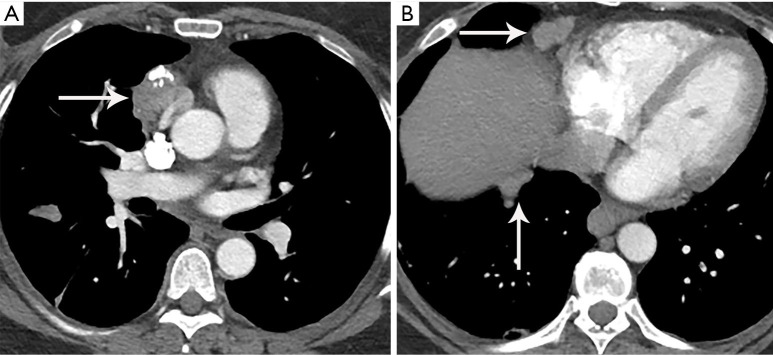
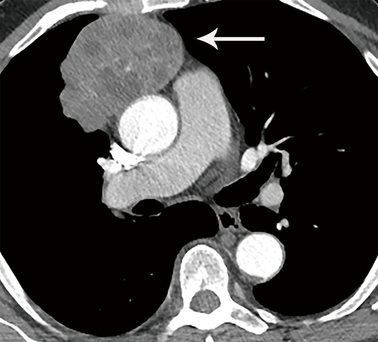
An integral part of managing patients with thymoma and thymic carcinoma is imaging. At diagnosis and staging, imaging helps demonstrate the extent of local invasion and distant metastases which allows the proper stratification of patients for therapy. For decades, the predominant staging system for thymic tumors was the Masaoka-Koga staging system. More recently, however, the International Association for the Study of Lung Cancer, the International Thymic Malignancies Interest Group (ITMIG), the European Society of Thoracic Surgeons, the Chinese Alliance for Research on Thymomas, and the Japanese Association of Research on Thymus partnered together to develop a tumor-node-metastasis (TNM) staging system specifically for thymic tumors based on a retrospective database of nearly 10,000 patients. The TNM 8 edition defines specific criteria for thymic tumors. Imaging also serves to assess treatment response and detect recurrent disease after various treatment modalities. The Response Evaluation Criteria in Solid Tumors (RECIST) version 1.1 is currently used to assess response to treatment. ITMIG recommends certain modifications to RECIST version 1.1, however, in thymic tumors due to unique patterns of spread. While there is often overlap, computed tomography (CT), magnetic resonance imaging (MRI), and positron emission tomography/computed tomography (PET/CT) characteristics can help differentiate thymoma and thymic carcinoma, with newer CT and MRI techniques under evaluation showing encouraging potential.
对胸腺瘤和胸腺癌患者进行管理的一个不可或缺的部分是影像学检查。在诊断和分期时,影像学有助于显示局部侵犯和远处转移的范围,从而使患者能够得到恰当的治疗分层。几十年来,胸腺肿瘤的主要分期系统是Masaoka-Koga分期系统。然而,最近,国际肺癌研究协会、国际胸腺恶性肿瘤研究组(ITMIG)、欧洲胸外科医师协会、中国胸腺瘤研究联盟和日本胸腺研究协会合作,基于近10000例患者的回顾性数据库,专门为胸腺肿瘤开发了一种肿瘤-淋巴结-转移(TNM)分期系统。TNM第8版定义了胸腺肿瘤的具体标准。影像学检查还用于评估治疗反应以及在各种治疗方式后检测疾病复发情况。实体瘤疗效评价标准(RECIST)第1.1版目前用于评估治疗反应。然而,由于胸腺肿瘤独特的扩散模式,ITMIG建议对RECIST第1.1版进行某些修改。虽然计算机断层扫描(CT)、磁共振成像(MRI)和正电子发射断层扫描/计算机断层扫描(PET/CT)的特征通常存在重叠,但它们有助于区分胸腺瘤和胸腺癌,正在评估的新型CT和MRI技术显示出令人鼓舞的潜力。