Rosskothen-Kuhl Nicole, Hildebrandt Heika, Birkenhäger Ralf, Illing Robert-Benjamin
Neurobiological Research Laboratory, Section for Clinical and Experimental Otology, University Medical Center Freiburg, Freiburg, Germany.
Molecular Biological Laboratory, Section for Clinical and Experimental Otology, University Medical Center Freiburg, Freiburg, Germany.
Front Cell Neurosci. 2018 Feb 22;12:43. doi: 10.3389/fncel.2018.00043. eCollection 2018.
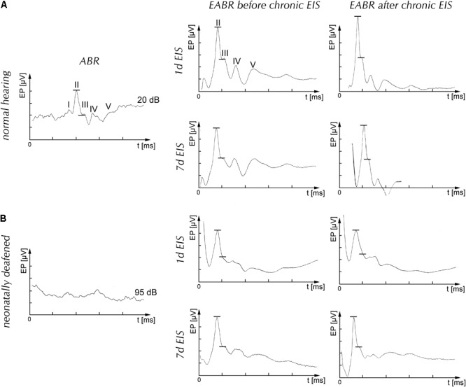
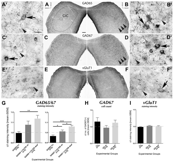
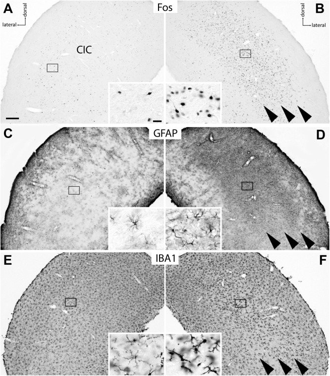
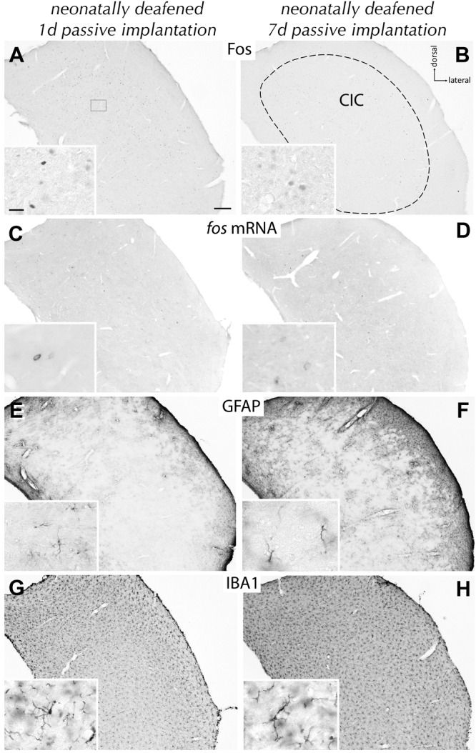
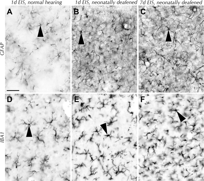
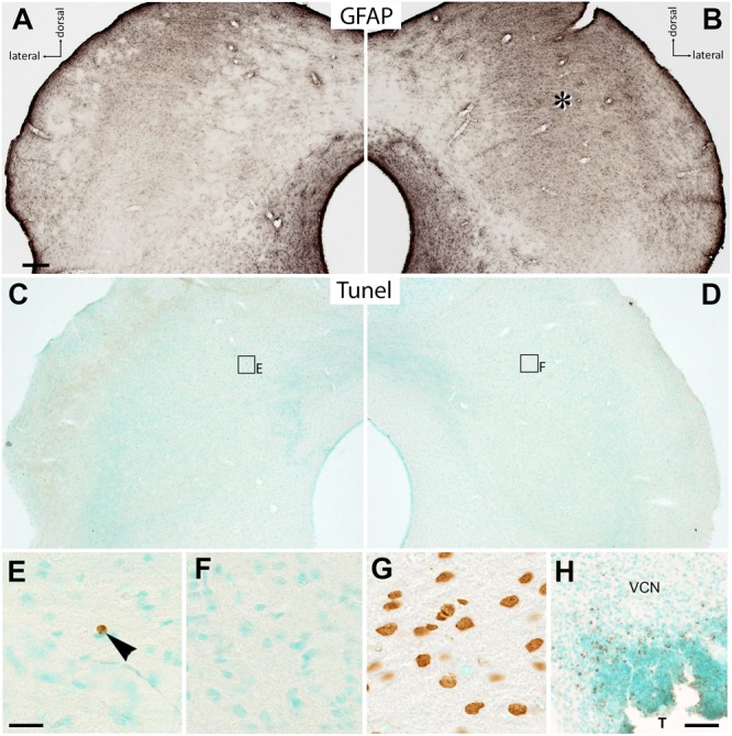
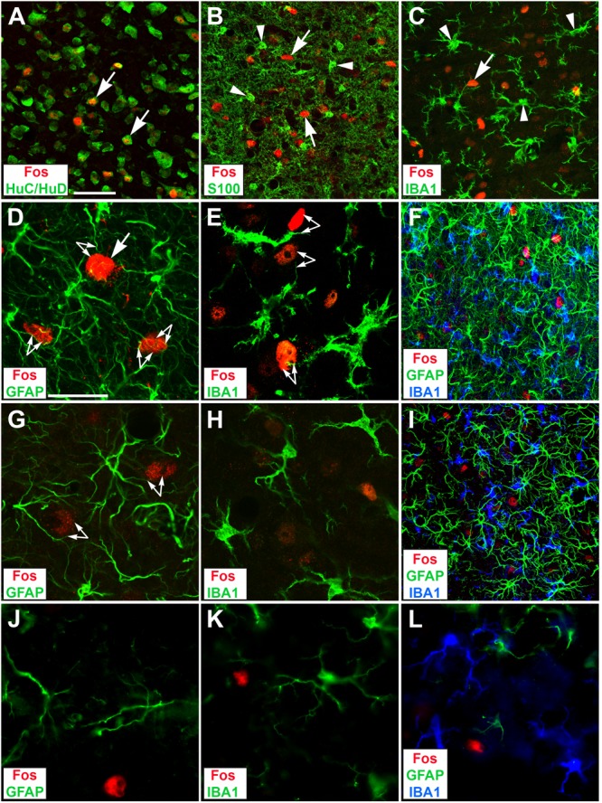
Neuron-glia interactions contribute to tissue homeostasis and functional plasticity in the mammalian brain, but it remains unclear how this is achieved. The potential of central auditory brain tissue for stimulation-dependent cellular remodeling was studied in hearing-experienced and neonatally deafened rats. At adulthood, both groups received an intracochlear electrode into the left cochlea and were continuously stimulated for 1 or 7 days after waking up from anesthesia. Normal hearing and deafness were assessed by auditory brainstem responses (ABRs). The effectiveness of stimulation was verified by electrically evoked ABRs as well as immunocytochemistry and hybridization for the immediate early gene product Fos on sections through the auditory midbrain containing the inferior colliculus (IC). Whereas hearing-experienced animals showed a tonotopically restricted Fos response in the IC contralateral to electrical intracochlear stimulation, Fos-positive neurons were found almost throughout the contralateral IC in deaf animals. In deaf rats, the Fos response was accompanied by a massive increase of GFAP indicating astrocytic hypertrophy, and a local activation of microglial cells identified by IBA1. These glia responses led to a noticeable increase of neuron-glia approximations. Moreover, staining for the GABA synthetizing enzymes GAD65 and GAD67 rose significantly in neuronal cell bodies and presynaptic boutons in the contralateral IC of deaf rats. Activation of neurons and glial cells and tissue re-composition were in no case accompanied by cell death as would have been apparent by a Tunel reaction. These findings suggest that growth and activity of glial cells is crucial for the local adjustment of neuronal inhibition to neuronal excitation.
神经元与神经胶质细胞的相互作用有助于维持哺乳动物大脑的组织稳态和功能可塑性,但目前尚不清楚这是如何实现的。我们在有听力经验的大鼠和新生致聋大鼠中研究了中枢听觉脑组织在刺激依赖性细胞重塑方面的潜力。成年后,两组大鼠的左耳蜗均植入了耳蜗内电极,并在从麻醉中苏醒后持续刺激1天或7天。通过听觉脑干反应(ABR)评估正常听力和耳聋情况。通过电诱发ABR以及免疫细胞化学和原位杂交技术检测包含下丘(IC)的听觉中脑切片上的即刻早期基因产物Fos,以验证刺激的有效性。有听力经验的动物在耳蜗内电刺激对侧的IC中表现出音调拓扑受限的Fos反应,而在致聋动物中,几乎在对侧IC的整个区域都发现了Fos阳性神经元。在致聋大鼠中,Fos反应伴随着GFAP的大量增加,表明星形胶质细胞肥大,以及由IBA1识别的小胶质细胞的局部激活。这些神经胶质细胞反应导致神经元与神经胶质细胞的接近度显著增加。此外,在致聋大鼠对侧IC的神经元细胞体和突触前终扣中,GABA合成酶GAD65和GAD67的染色显著增加。神经元和神经胶质细胞的激活以及组织重组在任何情况下都不会伴随着细胞死亡,否则Tunel反应会显示出来。这些发现表明,神经胶质细胞的生长和活性对于神经元抑制对神经元兴奋的局部调节至关重要。