Calza Giulio, Nyberg Elisabeth, Mäkinen Matias, Soliymani Rabah, Cascone Annunziata, Lindholm Dan, Barborini Emanuele, Baumann Marc, Lalowski Maciej, Eriksson Ove
Meilahti Clinical Proteomics Core Facility, Helsinki Institute of Life Science, Faculty of Medicine, University of Helsinki, Helsinki, Finland.
Department of Biochemistry and Developmental Biology, Faculty of Medicine, University of Helsinki, Helsinki, Finland.
Front Pharmacol. 2018 Feb 22;9:141. doi: 10.3389/fphar.2018.00141. eCollection 2018.
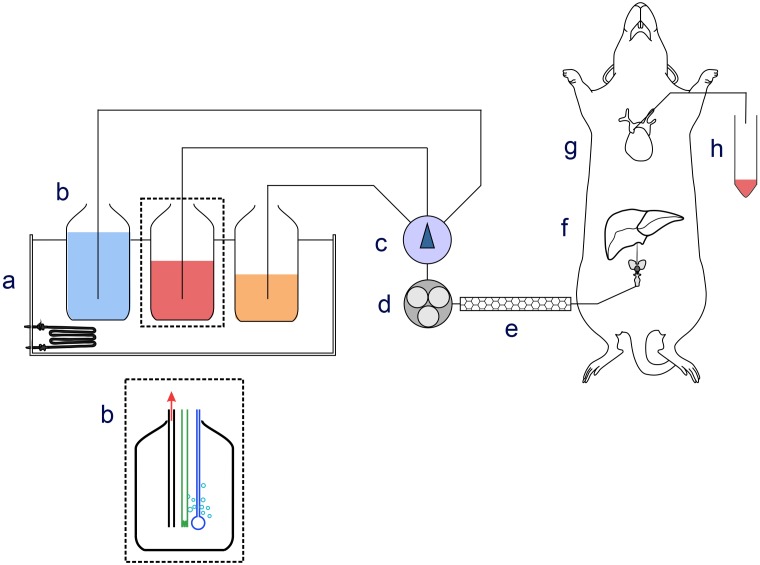
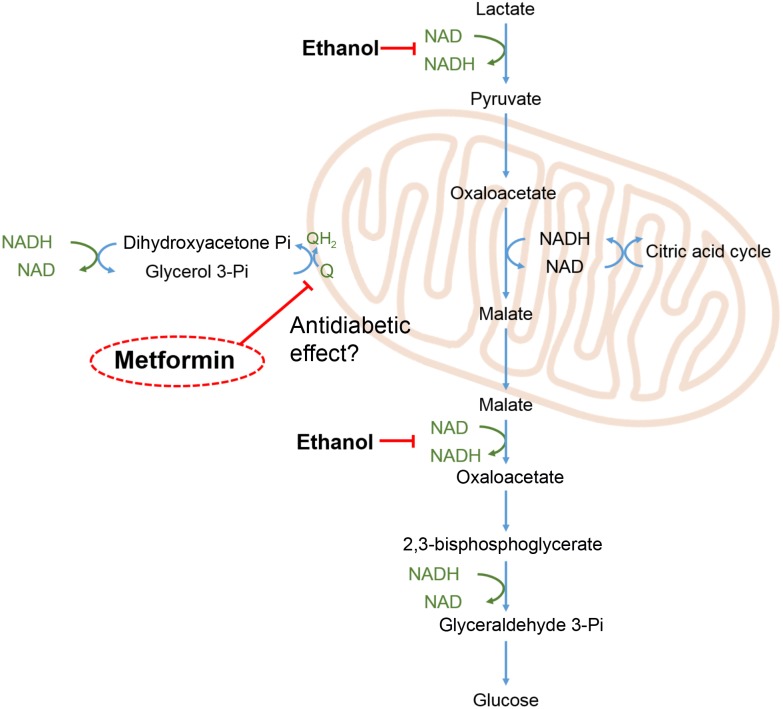
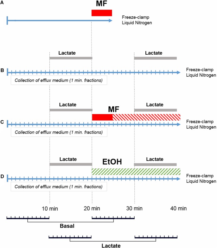
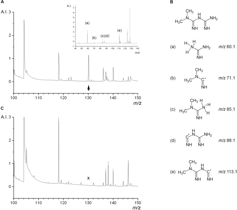
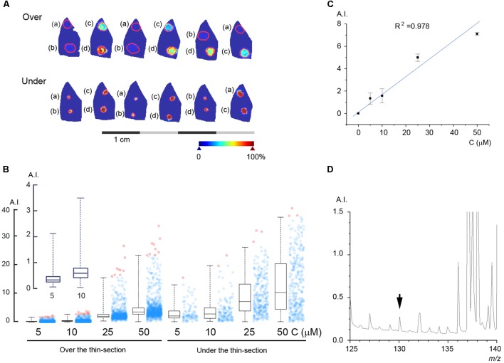
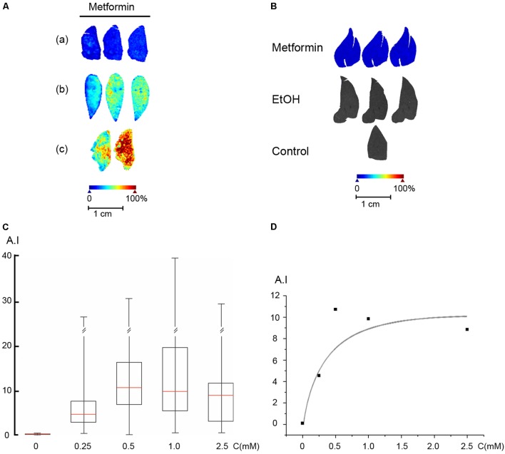
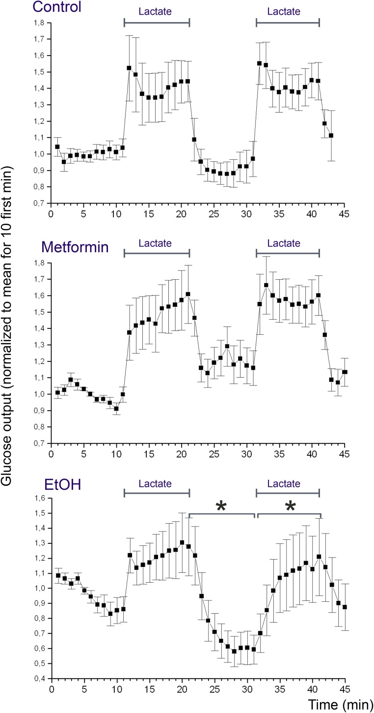
Metformin is the first line drug for type 2 diabetes but its molecular mechanisms remain unclear. Here, we have studied the acute effect of a therapeutically relevant intrahepatic concentration of metformin on glucose production from lactate. We selected the perfused rat liver as experimental system since it enables the complete control of drug dosage. We used MALDI (matrix-assisted laser desorption/ionization) mass spectrometry imaging to estimate the concentration of metformin in the livers and we measured the concentration of glucose in the effluent medium under basal conditions and following lactate addition. MALDI mass spectra of thin-sections of freeze-clamped rat liver perfused with metformin showed a peak at 130.16 which was unambiguously assigned to metformin. The mass spectrometric detection limit was at a tissue concentration of about 250 nM, and uptake of metformin from the perfusion medium to the liver occurred with a K of 0.44 mM. Metformin was evenly distributed in the liver irrespective of its concentration in the perfusion medium and the duration of a perfusion. At a parenchymal concentration of 30 μM, metformin did not induce any significant suppression of the basal or lactate-induced glucose release from the liver. These results show that matrix-assisted laser desorption/ionization mass spectrometry imaging can be applied to estimate the tissue concentration and distribution of metformin in a therapeutically relevant micromolar concentration range. Our findings challenge the view that metformin causes an inhibition of glucose release from the liver by an acute inhibition of mitochondrial glycerol 3-phosphate dehydrogenase (EC 1.1.5.3).
二甲双胍是2型糖尿病的一线用药,但其分子机制仍不清楚。在此,我们研究了治疗相关肝内浓度的二甲双胍对乳酸生成葡萄糖的急性作用。我们选择灌注大鼠肝脏作为实验系统,因为它能完全控制药物剂量。我们使用基质辅助激光解吸/电离(MALDI)质谱成像来估计肝脏中二甲双胍的浓度,并在基础条件下和添加乳酸后测量流出介质中葡萄糖的浓度。用二甲双胍灌注的冷冻钳夹大鼠肝脏薄片的MALDI质谱显示在130.16处有一个峰,明确归属于二甲双胍。质谱检测限为组织浓度约250 nM,二甲双胍从灌注介质到肝脏的摄取K为0.44 mM。无论其在灌注介质中的浓度和灌注持续时间如何,二甲双胍在肝脏中均均匀分布。在实质浓度为30 μM时,二甲双胍并未对基础或乳酸诱导的肝脏葡萄糖释放产生任何显著抑制作用。这些结果表明,基质辅助激光解吸/电离质谱成像可用于估计治疗相关微摩尔浓度范围内二甲双胍的组织浓度和分布。我们的发现对二甲双胍通过急性抑制线粒体甘油3-磷酸脱氢酶(EC 1.1.5.3)来抑制肝脏葡萄糖释放这一观点提出了挑战。