• 文献检索
  • 文档翻译
  • 深度研究
  • 学术资讯
  • Suppr Zotero 插件Zotero 插件
  • 邀请有礼
  • 套餐&价格
  • 历史记录
应用&插件
Suppr Zotero 插件Zotero 插件浏览器插件Mac 客户端Windows 客户端微信小程序
定价
高级版会员购买积分包购买API积分包
服务
文献检索文档翻译深度研究API 文档MCP 服务
关于我们
关于 Suppr公司介绍联系我们用户协议隐私条款
关注我们

Suppr 超能文献

核心技术专利:CN118964589B侵权必究
粤ICP备2023148730 号-1Suppr @ 2026

文献检索

告别复杂PubMed语法,用中文像聊天一样搜索,搜遍4000万医学文献。AI智能推荐,让科研检索更轻松。

立即免费搜索

文件翻译

保留排版,准确专业,支持PDF/Word/PPT等文件格式,支持 12+语言互译。

免费翻译文档

深度研究

AI帮你快速写综述,25分钟生成高质量综述,智能提取关键信息,辅助科研写作。

立即免费体验

通过滴注ROCK抑制剂力法舒地尔增加光学相干断层扫描血管造影术检测的视乳头周围血管密度:与溴莫尼定的比较

Increase in the OCT angiographic peripapillary vessel density by ROCK inhibitor ripasudil instillation: a comparison with brimonidine.

作者信息

Chihara Etsuo, Dimitrova Galina, Chihara Tomoyuki

机构信息

Sensho-kai Eye Institute, Minamiyama 50-1, Iseda, Uji, Kyoto, 611-0043, Japan.

Department of Ophthalmology, City General Hospital 8th September, Skopje, Macedonia.

出版信息

Graefes Arch Clin Exp Ophthalmol. 2018 Jul;256(7):1257-1264. doi: 10.1007/s00417-018-3945-5. Epub 2018 Mar 8.

DOI:10.1007/s00417-018-3945-5
PMID:29520478
原文链接:https://pmc.ncbi.nlm.nih.gov/articles/PMC6006239/
Abstract

PURPOSE

To assess the responses of the superficial peripapillary retinal vessel density (VD) and prelaminar flow index (PLFI) to topical Rho-assisted coiled-coil forming protein kinase (ROCK) inhibitor ripasudil and alpha-2 agonist brimonidine using optical coherence tomography angiography.

METHODS

This is a prospective, non-randomized, comparative cohort study. We studied the response of optical coherence tomography angiography (OCTA) parameters to drugs in 24 eyes treated with ripasudil and 23 eyes treated with brimonidine at the Sensho-kai Eye Institute. After division by the signal strength (SS), we compared the responses of peripapillary VD/SS and PLFI/unit area (UA)/SS to topical eye drops in eyes with primary open-angle glaucoma (POAG) and ocular hypertension (OH).

RESULTS

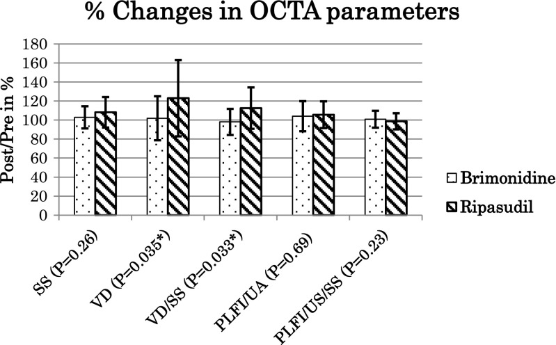
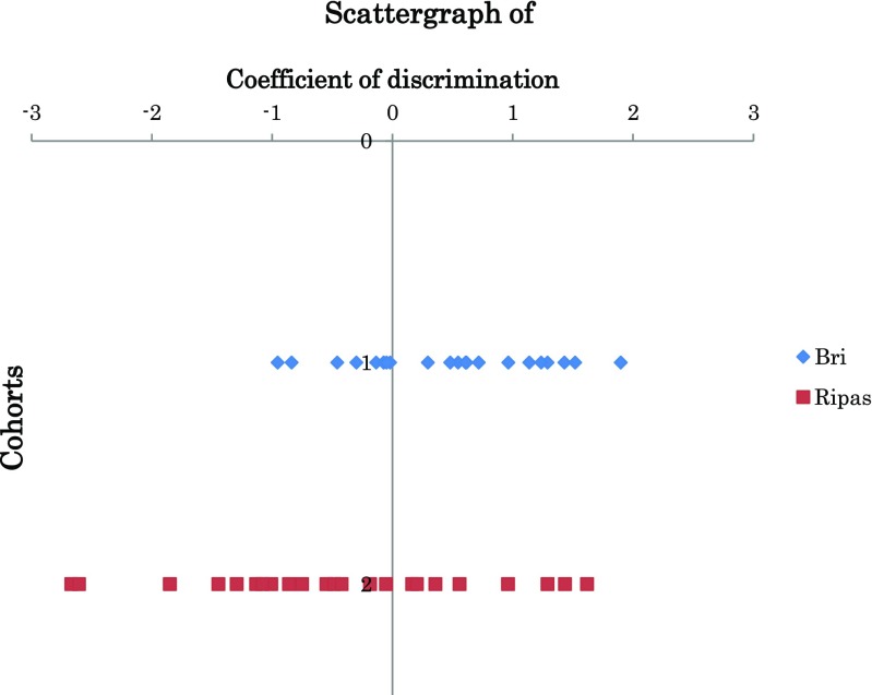
In the superficial peripapillary retina, VD/SS increased significantly in the ripasudil-treated eyes (12.5 ± 21.7%, P = 0.018), but not in the brimonidine-treated eyes (- 2.0 ± 13.8%, P = 0.484). In the deeper area of the optic disc, the changes in the PLFI/UA/SS in the brimonidine-treated eyes (+ 0.9 ± 8.9%, P = 1.00) and ripasudil-treated eyes (- 1.3 ± 8.5%, P = 0.241) were not significant. Multivariate discriminant analysis showed that the change in the peripapillary VD/SS was the most important parameter (P = 0.0186) for differentiating ripasudil- and brimonidine-treated eyes.

CONCLUSIONS

The topical ROCK inhibitor ripasudil enhanced the peripapillary VD in POAG and OH, whereas the alpha-2 agonist brimonidine did not. The PLFI did not respond to either drug.

摘要

目的

使用光学相干断层扫描血管造影术评估视乳头周围视网膜浅层血管密度(VD)和板层前血流指数(PLFI)对局部应用Rho辅助卷曲螺旋形成蛋白激酶(ROCK)抑制剂ripasudil和α-2激动剂溴莫尼定的反应。

方法

这是一项前瞻性、非随机、比较性队列研究。我们在Sensho-kai眼科研究所研究了光学相干断层扫描血管造影(OCTA)参数对24只接受ripasudil治疗的眼睛和23只接受溴莫尼定治疗的眼睛中药物的反应。按信号强度(SS)划分后,我们比较了原发性开角型青光眼(POAG)和高眼压症(OH)患者眼中视乳头周围VD/SS和PLFI/单位面积(UA)/SS对局部滴眼液的反应。

结果

在视乳头周围视网膜浅层,ripasudil治疗组的眼睛VD/SS显著增加(12.5±21.7%,P = 0.018),而溴莫尼定治疗组的眼睛则没有增加(-2.0±13.8%,P = 0.484)。在视盘较深区域,溴莫尼定治疗组眼睛(+0.9±8.9%,P = 1.00)和ripasudil治疗组眼睛(-1.3±8.5%,P = 0.241)的PLFI/UA/SS变化不显著。多变量判别分析表明,视乳头周围VD/SS的变化是区分ripasudil和溴莫尼定治疗组眼睛的最重要参数(P = 0.0186)。

结论

局部应用ROCK抑制剂ripasudil可提高POAG和OH患者视乳头周围的VD,而α-2激动剂溴莫尼定则不能。PLFI对两种药物均无反应。

https://cdn.ncbi.nlm.nih.gov/pmc/blobs/7035/6006239/9122b664b82c/417_2018_3945_Fig3_HTML.jpg
https://cdn.ncbi.nlm.nih.gov/pmc/blobs/7035/6006239/071f51097651/417_2018_3945_Fig1_HTML.jpg
https://cdn.ncbi.nlm.nih.gov/pmc/blobs/7035/6006239/8c756e3e61dd/417_2018_3945_Fig2_HTML.jpg
https://cdn.ncbi.nlm.nih.gov/pmc/blobs/7035/6006239/9122b664b82c/417_2018_3945_Fig3_HTML.jpg
https://cdn.ncbi.nlm.nih.gov/pmc/blobs/7035/6006239/071f51097651/417_2018_3945_Fig1_HTML.jpg
https://cdn.ncbi.nlm.nih.gov/pmc/blobs/7035/6006239/8c756e3e61dd/417_2018_3945_Fig2_HTML.jpg
https://cdn.ncbi.nlm.nih.gov/pmc/blobs/7035/6006239/9122b664b82c/417_2018_3945_Fig3_HTML.jpg

相似文献

1
Increase in the OCT angiographic peripapillary vessel density by ROCK inhibitor ripasudil instillation: a comparison with brimonidine.通过滴注ROCK抑制剂力法舒地尔增加光学相干断层扫描血管造影术检测的视乳头周围血管密度:与溴莫尼定的比较
Graefes Arch Clin Exp Ophthalmol. 2018 Jul;256(7):1257-1264. doi: 10.1007/s00417-018-3945-5. Epub 2018 Mar 8.
2
Discriminatory Power of Superficial Vessel Density and Prelaminar Vascular Flow Index in Eyes With Glaucoma and Ocular Hypertension and Normal Eyes.青光眼、高眼压症患者及正常眼的表层血管密度和板层前血管血流指数的鉴别能力
Invest Ophthalmol Vis Sci. 2017 Jan 1;58(1):690-697. doi: 10.1167/iovs.16-20709.
3
Optical Coherence Tomography Angiography Vessel Density Changes in Normal-tension Glaucoma Treated With Carteolol, Brimonidine, or Dorzolamide.正常眼压性青光眼经卡替洛尔、溴莫尼定或多佐胺治疗后的光学相干断层扫描血管密度变化。
J Glaucoma. 2021 Aug 1;30(8):690-696. doi: 10.1097/IJG.0000000000001859.
4
Influence of Removing the Large Retinal Vessels-related Effect on Peripapillary Vessel Density Progression Analysis in Glaucoma.青光眼中大视网膜血管相关效应去除对周边血管密度进展分析的影响。
J Glaucoma. 2018 Aug;27(8):e137-e139. doi: 10.1097/IJG.0000000000000990.
5
Effect of brimonidine on vascular density and imagej-derived flow index of optic nerve head and macula in primary open angle glaucoma.布林佐胺对原发性开角型青光眼视盘和黄斑血管密度及 ImageJ 衍生血流指数的影响。
Int Ophthalmol. 2024 Jul 4;44(1):311. doi: 10.1007/s10792-024-03221-7.
6
Peripapillary retinal vessel density in eyes with acute primary angle closure: an optical coherence tomography angiography study.急性原发性闭角型青光眼患者视乳头周围视网膜血管密度:一项光学相干断层扫描血管造影研究
Graefes Arch Clin Exp Ophthalmol. 2017 May;255(5):1013-1018. doi: 10.1007/s00417-017-3593-1. Epub 2017 Feb 4.
7
Repeatability of vessel density measurements of optical coherence tomography angiography in normal and glaucoma eyes.光学相干断层扫描血管造影术在正常眼和青光眼眼中血管密度测量的可重复性。
Br J Ophthalmol. 2018 Mar;102(3):352-357. doi: 10.1136/bjophthalmol-2017-310637. Epub 2017 Jul 24.
8
OCTA vessel density changes in the macular zone in glaucomatous eyes.青光眼患者眼部黄斑区光学相干断层扫描血管密度的变化
Graefes Arch Clin Exp Ophthalmol. 2018 Aug;256(8):1499-1508. doi: 10.1007/s00417-018-3965-1. Epub 2018 Apr 10.
9
Optical Coherence Tomography Angiography of Optic Disc in Eyes With Primary Open-angle Glaucoma and Normal-tension Glaucoma.原发性开角型青光眼和正常眼压性青光眼患者视盘的光学相干断层扫描血管造影。
J Glaucoma. 2019 Mar;28(3):243-251. doi: 10.1097/IJG.0000000000001184.
10
Diurnal Variations of Peripapillary and Macular Vessel Density in Glaucomatous Eyes Using Optical Coherence Tomography Angiography.利用光相干断层扫描血管造影术观察青光眼患者的眼周和黄斑血管密度的日变化。
J Glaucoma. 2018 Apr;27(4):336-341. doi: 10.1097/IJG.0000000000000914.

引用本文的文献

1
Effect of Ripasudil on the Change Rates of the Circumpapillary Retinal Nerve Fiber Layer Thickness in Patients With Primary Open-Angle Glaucoma.ripasudil对原发性开角型青光眼患者视盘周围视网膜神经纤维层厚度变化率的影响
J Ophthalmol. 2025 Jan 15;2025:2962982. doi: 10.1155/joph/2962982. eCollection 2025.
2
The impact of intraocular pressure on optical coherence tomography angiography: A review of current evidence.眼压对光学相干断层扫描血管造影的影响:当前证据综述
Saudi J Ophthalmol. 2024 Jan 3;38(2):144-151. doi: 10.4103/sjopt.sjopt_112_23. eCollection 2024 Apr-Jun.
3
Structure Function Relationships of Vessel Density and Retinal Nerve Fiber Layer Thickness in Early Glaucomatous Eyes With High Myopia.

本文引用的文献

1
Peripapillary and Macular Vessel Density in Patients with Glaucoma and Single-Hemifield Visual Field Defect.青光眼和单半视野视觉缺损患者的视乳头周围和黄斑区血管密度
Ophthalmology. 2017 May;124(5):709-719. doi: 10.1016/j.ophtha.2017.01.004. Epub 2017 Feb 10.
2
Discriminatory Power of Superficial Vessel Density and Prelaminar Vascular Flow Index in Eyes With Glaucoma and Ocular Hypertension and Normal Eyes.青光眼、高眼压症患者及正常眼的表层血管密度和板层前血管血流指数的鉴别能力
Invest Ophthalmol Vis Sci. 2017 Jan 1;58(1):690-697. doi: 10.1167/iovs.16-20709.
3
Quantitative Retinal Optical Coherence Tomography Angiography in Patients With Diabetes Without Diabetic Retinopathy.
高度近视早期青光眼患者血管密度与视网膜神经纤维层厚度的结构-功能关系。
Invest Ophthalmol Vis Sci. 2023 Apr 3;64(4):14. doi: 10.1167/iovs.64.4.14.
4
Effects of Smoking on Optic Nerve Head Microvasculature Density in Glaucoma.吸烟对青光眼视神经头微血管密度的影响。
J Glaucoma. 2022 Sep 1;31(9):710-716. doi: 10.1097/IJG.0000000000002081. Epub 2022 Jul 19.
5
Longitudinal Structure-Function Relationship between Macular Vessel Density and Thickness and Central Visual Field in Early Glaucoma.早期青光眼黄斑血管密度和厚度与中心视野的纵向结构-功能关系。
Ophthalmol Glaucoma. 2022 Nov-Dec;5(6):648-657. doi: 10.1016/j.ogla.2022.06.004. Epub 2022 Jun 13.
6
Comparison of the Effects of Latanoprostene Bunod and Timolol on Retinal Blood Vessel Density: A Randomized Clinical Trial.布诺地前列素与噻吗洛尔对视网膜血管密度影响的比较:一项随机临床试验。
Am J Ophthalmol. 2022 Sep;241:120-129. doi: 10.1016/j.ajo.2022.04.022. Epub 2022 May 6.
7
Comparative Assessment of Retinal Blood Flow Velocity Changes Following Brimonidine and Brinzolamide Administration Using Retinal Function Imaging.应用视网膜功能成像对布林佐胺和溴莫尼定滴眼后视网膜血流速度变化的比较评估。
Transl Vis Sci Technol. 2022 Feb 1;11(2):1. doi: 10.1167/tvst.11.2.1.
8
Combined wide-field optical coherence tomography angiography density map for high myopic glaucoma detection.用于高度近视性青光眼检测的宽视野光学相干断层扫描血管造影密度图的联合应用。
Sci Rep. 2021 Nov 11;11(1):22034. doi: 10.1038/s41598-021-01661-0.
9
Rates of Circumpapillary Retinal Nerve Fiber Layer Thinning and Capillary Density Loss in Glaucomatous Eyes with Disc Hemorrhage.盘沿视网膜神经纤维层变薄和伴有盘出血的青光眼眼毛细血管密度丧失的比率。
Am J Ophthalmol. 2022 Mar;235:24-31. doi: 10.1016/j.ajo.2021.09.013. Epub 2021 Sep 26.
10
Optical coherence tomographic angiography study of perfusion recovery after surgical lowering of intraocular pressure.手术降低眼内压后灌注恢复的光学相干断层扫描血管造影研究。
Sci Rep. 2021 Aug 26;11(1):17251. doi: 10.1038/s41598-021-96225-7.
糖尿病性视网膜病变未累及者的定量视网膜光学相干断层扫描血管造影
Invest Ophthalmol Vis Sci. 2017 Jan 1;58(1):190-196. doi: 10.1167/iovs.16-20531.
4
OCT Angiography of the Peripapillary Retina in Primary Open-Angle Glaucoma.原发性开角型青光眼视乳头周围视网膜的光学相干断层扫描血管造影
Invest Ophthalmol Vis Sci. 2016 Nov 1;57(14):6265-6270. doi: 10.1167/iovs.16-20287.
5
Ocular Penetration and Pharmacokinetics of Ripasudil Following Topical Administration to Rabbits.兔局部应用力法舒地尔的眼内通透性和药代动力学
J Ocul Pharmacol Ther. 2016 Sep;32(7):405-14. doi: 10.1089/jop.2016.0028. Epub 2016 Jul 27.
6
Projection-resolved optical coherence tomographic angiography.投影分辨光学相干断层扫描血管造影术
Biomed Opt Express. 2016 Feb 9;7(3):816-28. doi: 10.1364/BOE.7.000816. eCollection 2016 Mar 1.
7
Optic Disc Perfusion in Primary Open Angle and Normal Tension Glaucoma Eyes Using Optical Coherence Tomography-Based Microangiography.使用基于光学相干断层扫描的微血管造影术评估原发性开角型青光眼和正常眼压性青光眼患者视盘的血流灌注情况
PLoS One. 2016 May 5;11(5):e0154691. doi: 10.1371/journal.pone.0154691. eCollection 2016.
8
Vascular Normalization by ROCK Inhibitor: Therapeutic Potential of Ripasudil (K-115) Eye Drop in Retinal Angiogenesis and Hypoxia.ROCK抑制剂介导的血管正常化:玻璃体内注射法舒地尔(K-115)对视网膜血管生成及缺氧的治疗潜力
Invest Ophthalmol Vis Sci. 2016 Apr 1;57(4):2264-76. doi: 10.1167/iovs.15-17411.
9
Ultrahigh-Speed, Swept-Source Optical Coherence Tomography Angiography in Nonexudative Age-Related Macular Degeneration with Geographic Atrophy.超高速度、扫频源光学相干断层扫描血管造影在伴有地图样萎缩的非渗出性年龄相关性黄斑变性中的应用
Ophthalmology. 2015 Dec;122(12):2532-44. doi: 10.1016/j.ophtha.2015.08.029. Epub 2015 Oct 17.
10
Optical Coherence Tomography Angiography of the Peripapillary Retina in Glaucoma.青光眼患者视乳头周围视网膜的光学相干断层扫描血管造影
JAMA Ophthalmol. 2015 Sep;133(9):1045-52. doi: 10.1001/jamaophthalmol.2015.2225.