• 文献检索
  • 文档翻译
  • 深度研究
  • 学术资讯
  • Suppr Zotero 插件Zotero 插件
  • 邀请有礼
  • 套餐&价格
  • 历史记录
应用&插件
Suppr Zotero 插件Zotero 插件浏览器插件Mac 客户端Windows 客户端微信小程序
定价
高级版会员购买积分包购买API积分包
服务
文献检索文档翻译深度研究API 文档MCP 服务
关于我们
关于 Suppr公司介绍联系我们用户协议隐私条款
关注我们

Suppr 超能文献

核心技术专利:CN118964589B侵权必究
粤ICP备2023148730 号-1Suppr @ 2026

文献检索

告别复杂PubMed语法,用中文像聊天一样搜索,搜遍4000万医学文献。AI智能推荐,让科研检索更轻松。

立即免费搜索

文件翻译

保留排版,准确专业,支持PDF/Word/PPT等文件格式,支持 12+语言互译。

免费翻译文档

深度研究

AI帮你快速写综述,25分钟生成高质量综述,智能提取关键信息,辅助科研写作。

立即免费体验

旁分泌细胞衰老加剧胆管损伤并损害再生。

Paracrine cellular senescence exacerbates biliary injury and impairs regeneration.

机构信息

MRC Centre for Regenerative Medicine, University of Edinburgh, 5 Little France Drive, Edinburgh, EH16 4UU, UK.

MRC Centre for Inflammation Research, University of Edinburgh, Edinburgh, EH16 4TJ, UK.

出版信息

Nat Commun. 2018 Mar 9;9(1):1020. doi: 10.1038/s41467-018-03299-5.

DOI:10.1038/s41467-018-03299-5
PMID:29523787
原文链接:https://pmc.ncbi.nlm.nih.gov/articles/PMC5844882/
Abstract

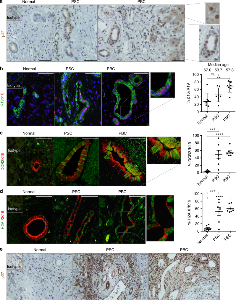
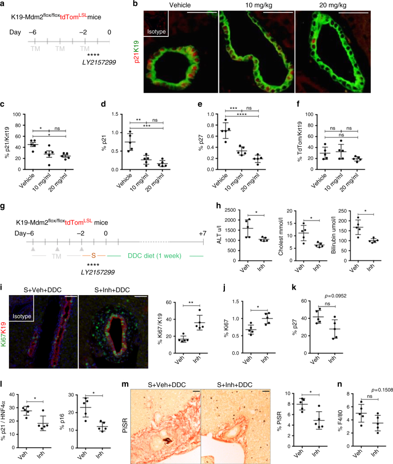
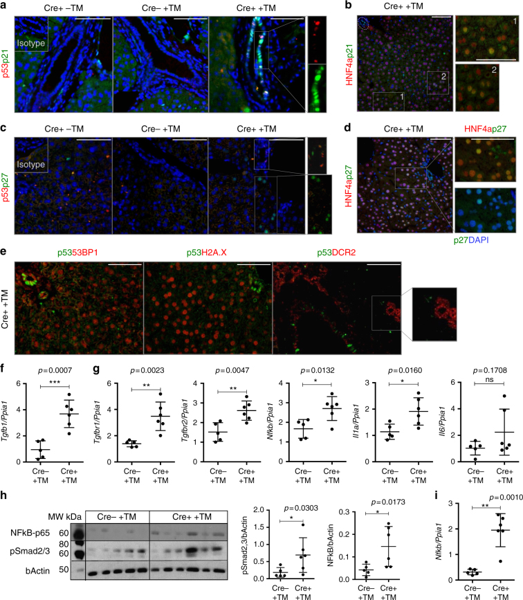
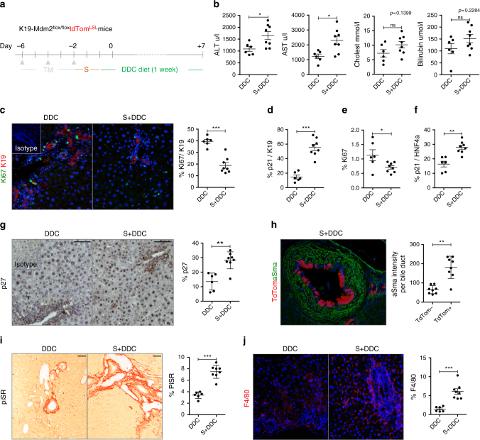
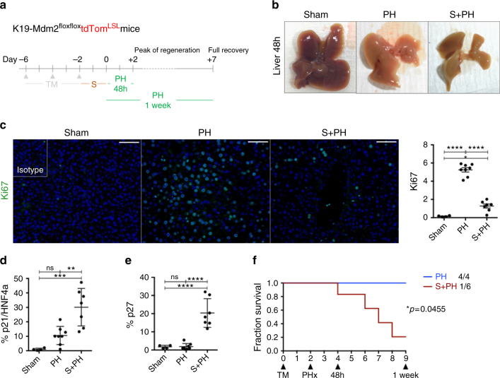
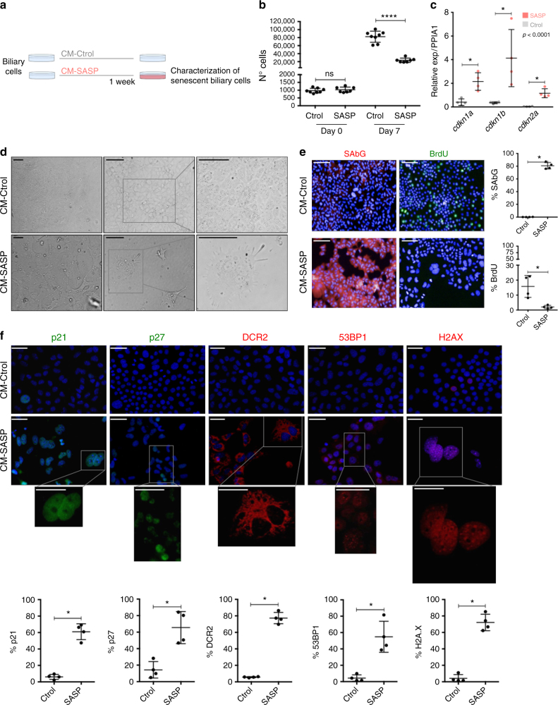
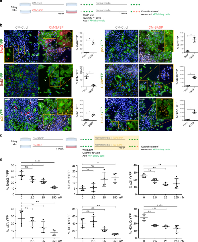
Cellular senescence is a mechanism that provides an irreversible barrier to cell cycle progression to prevent undesired proliferation. However, under pathological circumstances, senescence can adversely affect organ function, viability and regeneration. We have developed a mouse model of biliary senescence, based on the conditional deletion of Mdm2 in bile ducts under the control of the Krt19 promoter, that exhibits features of biliary disease. Here we report that senescent cholangiocytes induce profound alterations in the cellular and signalling microenvironment, with recruitment of myofibroblasts and macrophages causing collagen deposition, TGFβ production and induction of senescence in surrounding cholangiocytes and hepatocytes. Finally, we study how inhibition of TGFβ-signalling disrupts the transmission of senescence and restores liver function. We identify cellular senescence as a detrimental mechanism in the development of biliary injury. Our results identify TGFβ as a potential therapeutic target to limit senescence-dependent aggravation in human cholangiopathies.

摘要

细胞衰老(cellular senescence)是一种提供细胞周期进程不可逆屏障的机制,以防止不必要的增殖。然而,在病理情况下,衰老会对器官功能、活力和再生产生不利影响。我们开发了一种基于 Krt19 启动子控制下胆管中 Mdm2 条件性缺失的胆汁性衰老小鼠模型,该模型表现出胆汁性疾病的特征。在这里,我们报告称,衰老的胆管细胞诱导细胞和信号微环境发生深刻变化,招募肌成纤维细胞和巨噬细胞导致胶原沉积、TGFβ 产生,并诱导周围胆管细胞和肝细胞衰老。最后,我们研究了如何抑制 TGFβ 信号转导破坏衰老的传递并恢复肝功能。我们将细胞衰老确定为胆汁性损伤发展中的一种有害机制。我们的结果表明,TGFβ 是限制人类胆管病中与衰老相关的恶化的潜在治疗靶点。

https://cdn.ncbi.nlm.nih.gov/pmc/blobs/ec8f/5844882/aacd1ddd7f62/41467_2018_3299_Fig8_HTML.jpg
https://cdn.ncbi.nlm.nih.gov/pmc/blobs/ec8f/5844882/522668e07020/41467_2018_3299_Fig1_HTML.jpg
https://cdn.ncbi.nlm.nih.gov/pmc/blobs/ec8f/5844882/e374d527fc22/41467_2018_3299_Fig2_HTML.jpg
https://cdn.ncbi.nlm.nih.gov/pmc/blobs/ec8f/5844882/38ff5ce7d2bf/41467_2018_3299_Fig3_HTML.jpg
https://cdn.ncbi.nlm.nih.gov/pmc/blobs/ec8f/5844882/7910e5f22e47/41467_2018_3299_Fig4_HTML.jpg
https://cdn.ncbi.nlm.nih.gov/pmc/blobs/ec8f/5844882/7a0bcbe87289/41467_2018_3299_Fig5_HTML.jpg
https://cdn.ncbi.nlm.nih.gov/pmc/blobs/ec8f/5844882/08893696ab5c/41467_2018_3299_Fig6_HTML.jpg
https://cdn.ncbi.nlm.nih.gov/pmc/blobs/ec8f/5844882/dce65e121685/41467_2018_3299_Fig7_HTML.jpg
https://cdn.ncbi.nlm.nih.gov/pmc/blobs/ec8f/5844882/aacd1ddd7f62/41467_2018_3299_Fig8_HTML.jpg

相似文献

1
Paracrine cellular senescence exacerbates biliary injury and impairs regeneration.旁分泌细胞衰老加剧胆管损伤并损害再生。
Nat Commun. 2018 Mar 9;9(1):1020. doi: 10.1038/s41467-018-03299-5.
2
Chronic cholestatic liver diseases: clues from histopathology for pathogenesis.慢性胆汁淤积性肝病:从组织病理学角度探讨其发病机制
Mol Aspects Med. 2014 Jun;37:35-56. doi: 10.1016/j.mam.2013.10.001. Epub 2013 Oct 17.
3
Knockout of secretin receptor reduces biliary damage and liver fibrosis in Mdr2 mice by diminishing senescence of cholangiocytes.敲除分泌素受体可通过减少胆管细胞衰老来减少 Mdr2 小鼠的胆汁淤积性损伤和肝纤维化。
Lab Invest. 2018 Nov;98(11):1449-1464. doi: 10.1038/s41374-018-0093-9. Epub 2018 Jul 5.
4
Paracrine modulation of cholangiocyte serotonin synthesis orchestrates biliary remodeling in adults.旁分泌调节胆管细胞 5-羟色胺合成协调成人胆管重塑。
Am J Physiol Gastrointest Liver Physiol. 2011 Feb;300(2):G303-15. doi: 10.1152/ajpgi.00368.2010. Epub 2010 Nov 11.
5
Autophagy mediates the process of cellular senescence characterizing bile duct damages in primary biliary cirrhosis.自噬介导了原发性胆汁性肝硬化中胆管损伤的细胞衰老过程。
Lab Invest. 2010 Jun;90(6):835-43. doi: 10.1038/labinvest.2010.56. Epub 2010 Mar 8.
6
Cellular senescence in the cholangiopathies: a driver of immunopathology and a novel therapeutic target.胆管病中的细胞衰老:免疫病理学的驱动因素和新的治疗靶点。
Semin Immunopathol. 2022 Jul;44(4):527-544. doi: 10.1007/s00281-022-00909-9. Epub 2022 Feb 17.
7
TGFβ inhibition restores a regenerative response in acute liver injury by suppressing paracrine senescence.TGFβ 抑制通过抑制旁分泌衰老恢复急性肝损伤的再生反应。
Sci Transl Med. 2018 Aug 15;10(454). doi: 10.1126/scitranslmed.aan1230.
8
Knockout of the Tachykinin Receptor 1 in the Mdr2 (Abcb4) Mouse Model of Primary Sclerosing Cholangitis Reduces Biliary Damage and Liver Fibrosis.原发性硬化性胆管炎 Mdr2(Abcb4)小鼠模型中速激肽受体 1 的敲除可减轻胆管损伤和肝纤维化。
Am J Pathol. 2020 Nov;190(11):2251-2266. doi: 10.1016/j.ajpath.2020.07.007. Epub 2020 Jul 23.
9
Development and characterization of cholangioids from normal and diseased human cholangiocytes as an in vitro model to study primary sclerosing cholangitis.从正常和患病的人胆管细胞中培养类胆管并进行表征,以此作为研究原发性硬化性胆管炎的体外模型。
Lab Invest. 2017 Nov;97(11):1385-1396. doi: 10.1038/labinvest.2017.63. Epub 2017 Sep 11.
10
Cholangiocytes in the pathogenesis of primary sclerosing cholangitis and development of cholangiocarcinoma.原发性硬化性胆管炎发病机制和胆管癌发生中的胆管细胞。
Biochim Biophys Acta Mol Basis Dis. 2018 Apr;1864(4 Pt B):1390-1400. doi: 10.1016/j.bbadis.2017.08.020. Epub 2017 Aug 25.

引用本文的文献

1
Redefining senescence through hepatocyte fate changes in liver diseases.通过肝脏疾病中肝细胞命运变化重新定义衰老。
Trends Endocrinol Metab. 2025 Aug 28. doi: 10.1016/j.tem.2025.08.003.
2
M2 macrophages promote PKM2 production in fibroblasts to alleviate UVB-induced photoaging.M2巨噬细胞促进成纤维细胞中PKM2的产生,以减轻紫外线B诱导的光老化。
Cell Cycle. 2025 Mar-Apr;24(5-8):86-102. doi: 10.1080/15384101.2025.2514988. Epub 2025 Jun 5.
3
Consensus classification of biliary complications after liver transplantation: guidelines from the BileducTx meeting.

本文引用的文献

1
The senescence-associated secretory phenotype induces cellular plasticity and tissue regeneration.衰老相关分泌表型诱导细胞可塑性和组织再生。
Genes Dev. 2017 Jan 15;31(2):172-183. doi: 10.1101/gad.290635.116. Epub 2017 Jan 31.
2
Tissue damage and senescence provide critical signals for cellular reprogramming in vivo.组织损伤和衰老为体内细胞重编程提供了关键信号。
Science. 2016 Nov 25;354(6315). doi: 10.1126/science.aaf4445.
3
NOTCH1 mediates a switch between two distinct secretomes during senescence.NOTCH1在衰老过程中介导两种不同分泌组之间的转换。
肝移植术后胆道并发症的共识分类:BileducTx会议指南
Br J Surg. 2025 Apr 30;112(5). doi: 10.1093/bjs/znae321.
4
Longitudinal paired liver biopsies and transcriptome profiling in alcohol-associated hepatitis reveal dynamic changes in cellular senescence.酒精性肝炎患者的纵向配对肝活检及转录组分析揭示了细胞衰老的动态变化。
Gut. 2025 Aug 7;74(9):1500-1513. doi: 10.1136/gutjnl-2024-334094.
5
Hepatocellular senescence induces multi-organ senescence and dysfunction via TGFβ.肝细胞衰老通过转化生长因子β(TGFβ)诱导多器官衰老和功能障碍。
Nat Cell Biol. 2024 Dec;26(12):2075-2083. doi: 10.1038/s41556-024-01543-3. Epub 2024 Nov 13.
6
Liver Macrophage Diversity in Health and Disease.肝脏巨噬细胞在健康与疾病中的多样性。
Results Probl Cell Differ. 2024;74:175-209. doi: 10.1007/978-3-031-65944-7_7.
7
Utilising an in silico model to predict outcomes in senescence-driven acute liver injury.利用计算机模拟模型预测衰老驱动的急性肝损伤的结果。
NPJ Regen Med. 2024 Sep 30;9(1):26. doi: 10.1038/s41536-024-00371-1.
8
Regulation of intestinal senescence during cholestatic liver disease modulates barrier function and liver disease progression.胆汁淤积性肝病期间肠道衰老的调节可调节屏障功能和肝病进展。
JHEP Rep. 2024 Jun 29;6(10):101159. doi: 10.1016/j.jhepr.2024.101159. eCollection 2024 Oct.
9
Central role for cholangiocyte pathobiology in cholestatic liver diseases.胆管细胞病理生物学在胆汁淤积性肝病中的核心作用。
Hepatology. 2024 Sep 9. doi: 10.1097/HEP.0000000000001093.
10
Targeting SIRT2 in Aging-Associated Fibrosis Pathophysiology.针对衰老相关纤维化病理生理学中的沉默调节蛋白2(SIRT2)
Aging Dis. 2024 Aug 15. doi: 10.14336/AD.202.0513.
Nat Cell Biol. 2016 Sep;18(9):979-92. doi: 10.1038/ncb3397. Epub 2016 Aug 15.
4
Clinical development of galunisertib (LY2157299 monohydrate), a small molecule inhibitor of transforming growth factor-beta signaling pathway.转化生长因子-β信号通路小分子抑制剂加鲁尼西替(LY2157299一水合物)的临床开发
Drug Des Devel Ther. 2015 Aug 10;9:4479-99. doi: 10.2147/DDDT.S86621. eCollection 2015.
5
Hepatic progenitor cells of biliary origin with liver repopulation capacity.具有肝脏再填充能力的胆管源性肝祖细胞。
Nat Cell Biol. 2015 Aug;17(8):971-983. doi: 10.1038/ncb3203. Epub 2015 Jul 20.
6
Absence of the intestinal microbiota exacerbates hepatobiliary disease in a murine model of primary sclerosing cholangitis.在原发性硬化性胆管炎的小鼠模型中,肠道微生物群的缺失会加剧肝胆疾病。
Hepatology. 2016 Jan;63(1):185-96. doi: 10.1002/hep.27927. Epub 2015 Aug 10.
7
Functional role of cellular senescence in biliary injury.细胞衰老在胆汁损伤中的功能作用。
Am J Pathol. 2015 Mar;185(3):602-9. doi: 10.1016/j.ajpath.2014.10.027. Epub 2015 Jan 22.
8
Cellular senescence: from physiology to pathology.细胞衰老:从生理学到病理学。
Nat Rev Mol Cell Biol. 2014 Jul;15(7):482-96. doi: 10.1038/nrm3823.
9
Cholangiocyte senescence by way of N-ras activation is a characteristic of primary sclerosing cholangitis.通过N-ras激活途径导致的胆管细胞衰老,是原发性硬化性胆管炎的一个特征。
Hepatology. 2014 Jun;59(6):2263-75. doi: 10.1002/hep.26993. Epub 2014 Apr 25.
10
Primary sclerosing cholangitis.原发性硬化性胆管炎。
Lancet. 2013 Nov 9;382(9904):1587-99. doi: 10.1016/S0140-6736(13)60096-3. Epub 2013 Jun 28.