Department of Medicine, Neurology Imaging Unit, Imperial College London, London, W12 0NN, UK.
Department of Neuroimaging, Institute of Psychiatry, Kings College London, London, WC2R 2LS, UK.
Eur J Nucl Med Mol Imaging. 2018 Jul;45(8):1432-1441. doi: 10.1007/s00259-018-3984-5. Epub 2018 Mar 9.
Neuroinflammation and microglial activation play an important role in amnestic mild cognitive impairment (MCI) and Alzheimer's disease. In this study, we investigated the spatial distribution of neuroinflammation in MCI subjects, using spectral analysis (SA) to generate parametric maps and quantify C-PBR28 PET, and compared these with compartmental and other kinetic models of quantification.
Thirteen MCI and nine healthy controls were enrolled in this study. Subjects underwent C-PBR28 PET scans with arterial cannulation. Spectral analysis with an arterial plasma input function was used to generate C-PBR28 parametric maps. These maps were then compared with regional C-PBR28 V (volume of distribution) using a two-tissue compartment model and Logan graphic analysis. Amyloid load was also assessed with F-Flutemetamol PET.
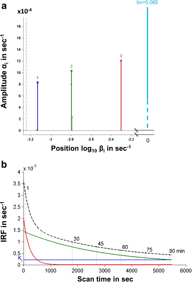
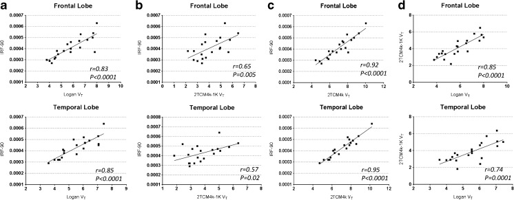
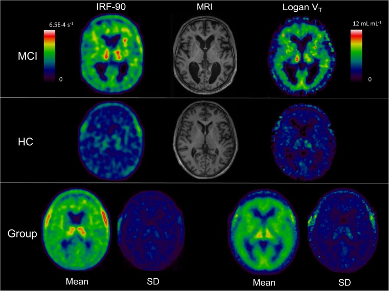
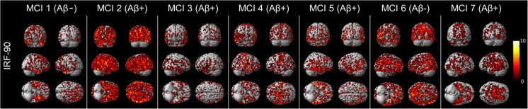
With SA, three component peaks were identified in addition to blood volume. The C-PBR28 impulse response function (IRF) at 90 min produced the lowest coefficient of variation. Single-subject analysis using this IRF demonstrated microglial activation in five out of seven amyloid-positive MCI subjects. IRF parametric maps of C-PBR28 uptake revealed a group-wise significant increase in neuroinflammation in amyloid-positive MCI subjects versus HC in multiple cortical association areas, and particularly in the temporal lobe. Interestingly, compartmental analysis detected group-wise increase in C-PBR28 binding in the thalamus of amyloid-positive MCI subjects, while Logan parametric maps did not perform well.
This study demonstrates for the first time that spectral analysis can be used to generate parametric maps of C-PBR28 uptake, and is able to detect microglial activation in amyloid-positive MCI subjects. IRF parametric maps of C-PBR28 uptake allow voxel-wise single-subject analysis and could be used to evaluate microglial activation in individual subjects.
神经炎症和小胶质细胞激活在遗忘型轻度认知障碍(MCI)和阿尔茨海默病中起着重要作用。在这项研究中,我们使用谱分析(SA)生成参数图并量化 C-PBR28 PET,来研究 MCI 患者的神经炎症的空间分布,并将其与定量的房室和其他动力学模型进行比较。
本研究纳入了 13 名 MCI 患者和 9 名健康对照者。受试者行 C-PBR28 PET 扫描并进行动脉插管。使用动脉血浆输入函数的谱分析生成 C-PBR28 参数图。然后,使用双室模型和 Logan 图形分析将这些参数图与区域 C-PBR28 V(分布容积)进行比较。还使用 F-Flutemetamol PET 评估淀粉样蛋白负荷。
通过 SA,除了血液体积外,还鉴定出三个组成峰。90 分钟时的 C-PBR28 脉冲响应函数(IRF)产生的变异系数最低。使用此 IRF 的单个体分析显示,在 7 名淀粉样蛋白阳性的 MCI 患者中有 5 名存在小胶质细胞激活。C-PBR28 摄取的 IRF 参数图显示,与 HC 相比,淀粉样蛋白阳性的 MCI 患者的多个皮质相关区域的神经炎症呈组间显著增加,尤其是在颞叶。有趣的是,房室分析检测到淀粉样蛋白阳性的 MCI 患者丘脑的 C-PBR28 结合呈组间增加,而 Logan 参数图表现不佳。
这项研究首次证明了谱分析可用于生成 C-PBR28 摄取的参数图,并能够检测淀粉样蛋白阳性的 MCI 患者中的小胶质细胞激活。C-PBR28 摄取的 IRF 参数图允许进行每个体素的单个体分析,并可用于评估个体受试者中的小胶质细胞激活。