Department of Molecular and Cell Biology, Henry M. Goldman School of Dental Medicine, Boston University, MA 02118, USA.
Institute of Cell Biology, University of Bern, Baltzerstrasse 4, CH-3012 Bern, Switzerland.
FEMS Microbiol Rev. 2018 May 1;42(3):324-334. doi: 10.1093/femsre/fuy007.
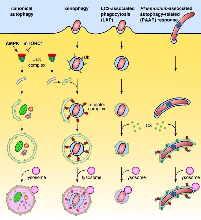
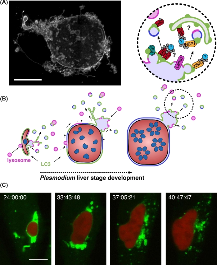
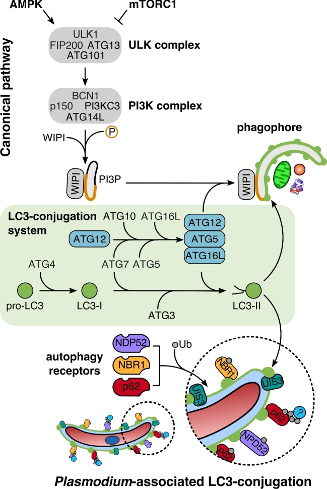
Recent years have witnessed a great gain in knowledge regarding parasite-host cell interactions during Plasmodium liver stage development. It is now an accepted fact that a large percentage of sporozoites invading hepatocytes fail to form infectious merozoites. There appears to be a delicate balance between parasite survival and elimination and we now start to understand why this is so. Plasmodium liver stage parasites replicate within the parasitophorous vacuole (PV), formed during invasion by invagination of the host cell plasma membrane. The main interface between the parasite and hepatocyte is the parasitophorous vacuole membrane (PVM) that surrounds the PV. Recently, it was shown that autophagy marker proteins decorate the PVM of Plasmodium liver stage parasites and eliminate a proportion of them by an autophagy-like mechanism. Successfully developing Plasmodium berghei parasites are initially also labeled but in the course of development, they are able to control this host defense mechanism by shedding PVM material into the tubovesicular network (TVN), an extension of the PVM that releases vesicles into the host cell cytoplasm. Better understanding of the molecular events at the PVM/TVN during parasite elimination could be the basis of new antimalarial measures.
近年来,人们对疟原虫肝脏阶段发育过程中寄生虫-宿主细胞相互作用的认识有了很大的提高。现在已经公认,很大一部分入侵肝细胞的子孢子无法形成感染性裂殖体。寄生虫的存活和消除之间似乎存在着微妙的平衡,我们现在开始理解为什么会这样。疟原虫在寄生泡(PV)内复制,寄生泡是在宿主细胞膜内陷入侵时形成的。寄生虫和肝细胞的主要界面是包围 PV 的寄生泡膜(PVM)。最近的研究表明,自噬标记蛋白装饰疟原虫肝脏阶段寄生虫的 PVM,并通过自噬样机制消除其中一部分。最初,发育良好的伯氏疟原虫也被标记,但在发育过程中,它们能够通过将 PVM 物质脱落到管状泡网络(TVN)中来控制这种宿主防御机制,TVN 是 PVM 的延伸,将囊泡释放到宿主细胞质中。更好地了解寄生虫消除过程中 PVM/TVN 的分子事件可能是新抗疟措施的基础。