Department of Respiratory Medicine, The Second People's Hospital of Longgang District, Shenzhen, China.
The Division of Pulmonary and Critical Care Medicine,The First Affiliated Hospital of Sun Yat-sen University; Institute of Respiratory Diseases of Sun Yat-sen University, Guanzhou, China.
Biosci Rep. 2018 Apr 27;38(2). doi: 10.1042/BSR20171555.
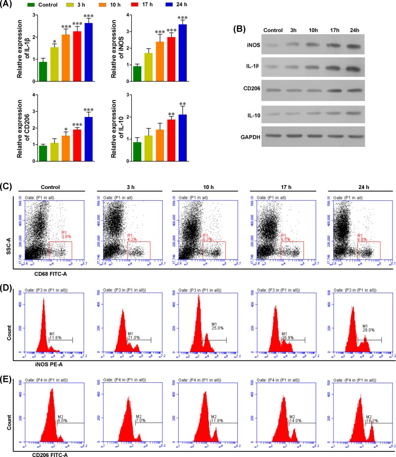
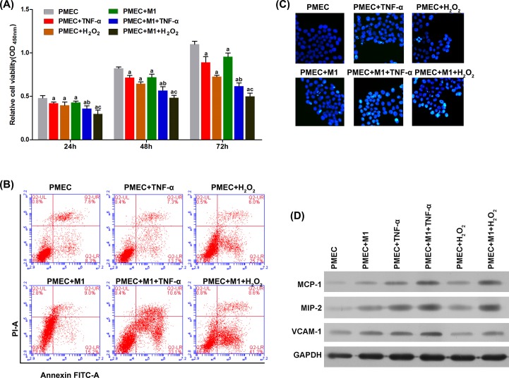
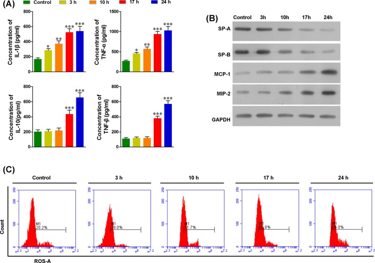
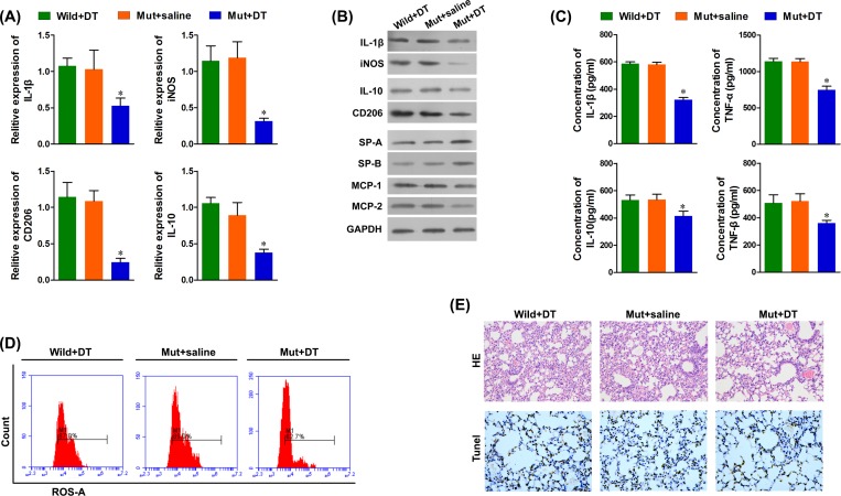
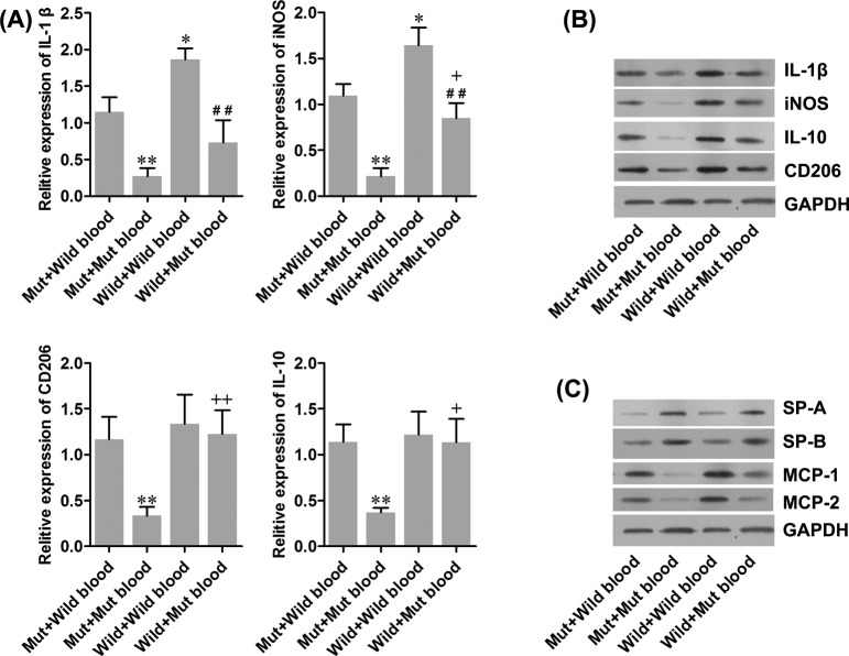
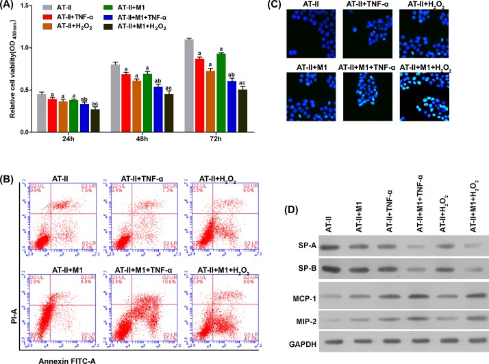
The goal of the present study was to investigate the role of M1 macrophages in acute lung injury (ALI). To address this, we used lipopolysaccharide (LPS)-treated wild-type and CD11b-DTR mice, and examined their M1 macrophage levels, and the extent of their inflammation and pulmonary injuries. In addition, we evaluated pulmonary function by measuring the expressions of SP-A and SP-B in infiltrated M1 macrophages. Finally, we co-cultured the mouse type II-like alveolar epithelial cells (AT-II) and mouse pulmonary microvascular endothelial cells (PMECs) with M1 macrophages in the presence of TNF-α or HO and assessed them for viability and apoptosis. After LPS treatment, we observed that the number of pulmonary M1/M2 macrophages and the serum levels of interleukin-1β (IL-1β), tumor necrosis factor α (TNF-α), and reactive oxygen species (ROS) significantly increased. Furthermore, the increase in cytokines was accompanied with the initiation of lung injury indicated by the decreased levels of SP-A and SP-B. In macrophage-depleted CD11b-DTR mice, ALI was attenuated, serum levels of IL-1β, TNF-α and ROS were reduced, and lung levels of monocyte chemoattractant protein-1 (MCP-1) and macrophage inflammatory protein-2 (MIP-2) were decreased. After administering TNF-α and HO, the proapoptotic effect of M1 macrophages on AT-II or PMECs significantly increased, the cell viabilities significantly decreased, and apoptosis significantly increased. Our results suggest that M1 macrophages are recruited to the lungs where they significantly contribute to an increase in TNF-α and ROS production, thus initiating ALI.
本研究旨在探讨 M1 巨噬细胞在急性肺损伤(ALI)中的作用。为此,我们使用脂多糖(LPS)处理野生型和 CD11b-DTR 小鼠,检测其 M1 巨噬细胞水平、炎症和肺损伤程度。此外,我们通过检测浸润 M1 巨噬细胞中 SP-A 和 SP-B 的表达来评估肺功能。最后,我们在 TNF-α或 HO 的存在下将小鼠 II 型肺泡上皮细胞(AT-II)和小鼠肺微血管内皮细胞(PMEC)与 M1 巨噬细胞共培养,并评估它们的活力和凋亡情况。在 LPS 处理后,我们观察到肺 M1/M2 巨噬细胞数量以及白细胞介素 1β(IL-1β)、肿瘤坏死因子 α(TNF-α)和活性氧(ROS)的血清水平显著增加。此外,细胞因子的增加伴随着肺损伤的开始,表现为 SP-A 和 SP-B 水平的降低。在巨噬细胞耗竭的 CD11b-DTR 小鼠中,ALI 得到缓解,IL-1β、TNF-α 和 ROS 的血清水平降低,肺单核细胞趋化蛋白-1(MCP-1)和巨噬细胞炎症蛋白-2(MIP-2)水平降低。给予 TNF-α和 HO 后,M1 巨噬细胞对 AT-II 或 PMEC 的促凋亡作用显著增强,细胞活力显著降低,凋亡显著增加。我们的结果表明,M1 巨噬细胞被募集到肺部,显著增加 TNF-α和 ROS 的产生,从而引发 ALI。