Department of Surgery, University Medical Center Groningen, Groningen, The Netherlands.
Institute for Medical and Biological Engineering, Schools of Engineering, Biological Sciences and Medicine, Pontificia Universidad Católica de Chile, Santiago, Chile.
Sci Rep. 2018 Mar 13;8(1):4405. doi: 10.1038/s41598-018-22689-9.
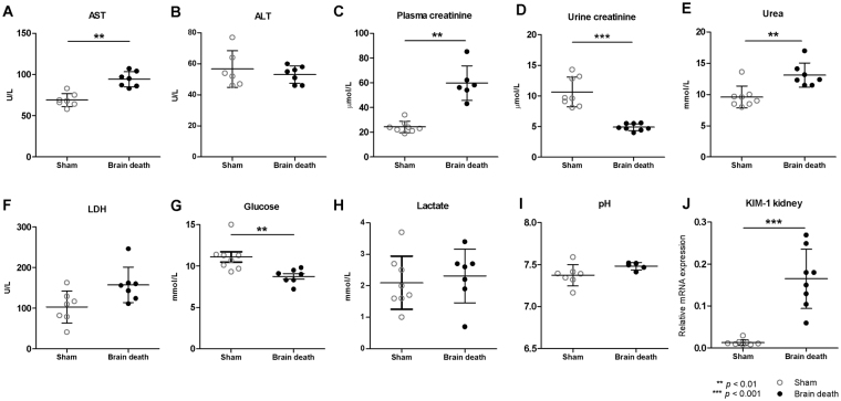
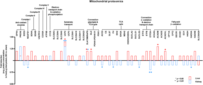
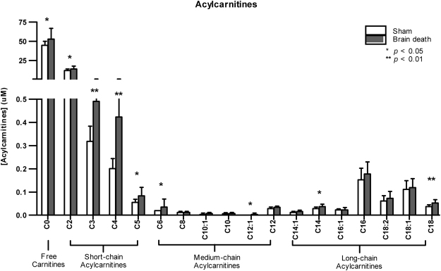
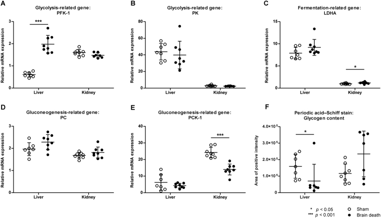
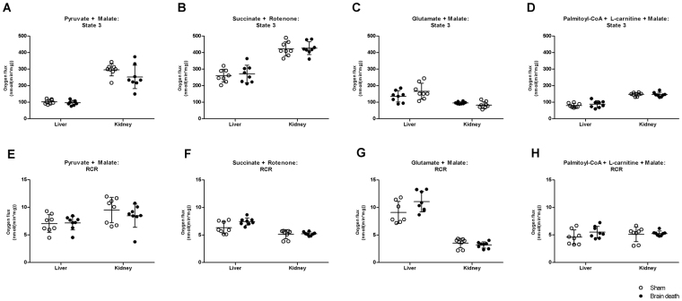
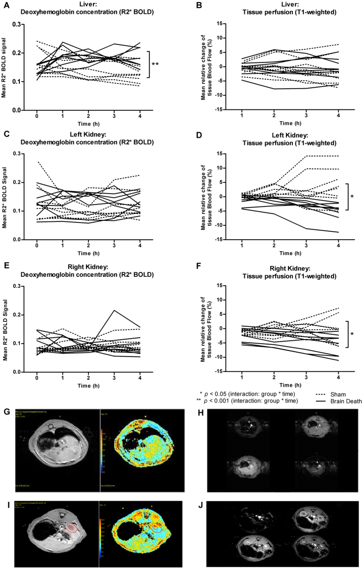
Hepatic and renal energy status prior to transplantation correlates with graft survival. However, effects of brain death (BD) on organ-specific energy status are largely unknown. We studied metabolism, perfusion, oxygen consumption, and mitochondrial function in the liver and kidneys following BD. BD was induced in mechanically-ventilated rats, inflating an epidurally-placed Fogarty-catheter, with sham-operated rats as controls. A 9.4T-preclinical MRI system measured hourly oxygen availability (BOLD-related R2*) and perfusion (T1-weighted). After 4 hrs, tissue was collected, mitochondria isolated and assessed with high-resolution respirometry. Quantitative proteomics, qPCR, and biochemistry was performed on stored tissue/plasma. Following BD, the liver increased glycolytic gene expression (Pfk-1) with decreased glycogen stores, while the kidneys increased anaerobic- (Ldha) and decreased gluconeogenic-related gene expression (Pck-1). Hepatic oxygen consumption increased, while renal perfusion decreased. ATP levels dropped in both organs while mitochondrial respiration and complex I/ATP synthase activity were unaffected. In conclusion, the liver responds to increased metabolic demands during BD, enhancing aerobic metabolism with functional mitochondria. The kidneys shift towards anaerobic energy production while renal perfusion decreases. Our findings highlight the need for an organ-specific approach to assess and optimise graft quality prior to transplantation, to optimise hepatic metabolic conditions and improve renal perfusion while supporting cellular detoxification.
移植前肝肾功能状态与移植物存活率相关。然而,脑死亡(BD)对器官特异性能量状态的影响尚不清楚。我们研究了 BD 后肝、肾的代谢、灌注、耗氧量和线粒体功能。使用机械通气大鼠,通过硬膜外放置的 Fogarty 导管充气诱导 BD,假手术大鼠作为对照。9.4T 临床前 MRI 系统每小时测量一次氧可用性(BOLD 相关 R2*)和灌注(T1 加权)。4 小时后,采集组织,分离线粒体,并使用高分辨率呼吸测定法进行评估。对储存的组织/血浆进行定量蛋白质组学、qPCR 和生物化学分析。BD 后,肝脏增加糖酵解基因表达(Pfk-1),同时糖原储存减少,而肾脏增加无氧(Ldha)并减少糖异生相关基因表达(Pck-1)。肝耗氧量增加,而肾灌注减少。两种器官的 ATP 水平下降,而线粒体呼吸和复合物 I/ATP 合酶活性不受影响。总之,肝脏在 BD 期间对代谢需求增加做出反应,通过功能线粒体增强有氧代谢。肾脏转向无氧能量产生,同时肾灌注减少。我们的研究结果强调了在移植前需要采用器官特异性方法评估和优化移植物质量,以优化肝代谢条件并改善肾灌注,同时支持细胞解毒。